The role of the epithelium and epithelial cytokines in eosinophilic esophagitis

The epithelium and epithelial cell-derived cytokines play an important role in the pathogenesis of eosinophilic esophagitis (EoE).1–3 Further understanding of EoE pathology may support biomarker identification and long-term disease control, and prevent disease progression4,5

EoE, when untreated, is a chronic and progressive inflammatory disorder of the oesophagus, characterised by inflammation, remodelling, and fibrosis1,2

  • EoE is associated with atopy, with 60–80% of patients with EoE found to have a history of atopic diseases, including asthma, atopic dermatitis, food allergies and allergic rhinitis.2,3 Environmental and genetic risk factors also play a key role in disease pathogenesis3–5
  • The epithelial barrier is disrupted in EoE, with basal zone hyperplasia, subepithelial fibrosis, immune cell infiltration, barrier impairment and smooth muscle hypertrophy observed5,6
  • Elevated expression of epithelial cell-derived cytokines has been observed in patients with EoE.7,8 Upon exposure to environmental insults, thymic stromal lymphopoietin (TSLP), interleukin (IL)-33 and IL‑25 are released, which drive multiple downstream inflammatory pathways;5,6,9 however, the role of IL-25 in EoE pathogenesis is less understood7
  • EoE diagnosis is often delayed, and reasons include but are not limited to: patients presenting with non-specific symptoms; adaptive eating behaviours; low awareness of EoE among healthcare professionals; and poor access and limited referrals to the correct care.10,11 Untreated disease may progress, with the risk of stricture increasing by 9% each year, highlighting the importance of limiting diagnostic delay12
  • EoE imposes a substantial burden on patients, caregivers and healthcare resources.1,13 Despite recent advances, there is an unmet need for treatments that control chronic inflammation and restore the epithelial barrier14–17

1. Lucendo AJ, et al. United European Gastroenterol J. 2017;5:335–358; 2. Dellon ES, et al. Am J Gastroenterol. 2025;120:31–59; 3. Jaros J, et al. J Clin Aesthetic Dermatol. 2025;18:15–20; 4. Muir A, et al. JAMA. 2021;326:1310–1318; 5. Canani BR, et al. Allergy. 2024;79:1485–1500; 6. Laky K, et al. J Allergy Clin Immunol. 2024;153:1485–1499; 7. Simon D, et al. Allergy. 2015;70:433–452; 8. Travers J, et al. Sci Rep. 2017;7:17563; 9. Farah A, et al. Diagnostics (Basel). 2025;15:240; 10. Muir AB, et al. Clin Exp Gastroenterol. 2019;12:391–399; 11. Kanakala V, et al. Eur J Gastroenterol Hepatol. 2010;22:848–85513; 12. Warners MJ, et al. Am J Gastroenterol. 2018;113:836–844; 13. Mukkada V, et al. Clin Gastroenterol Hepatol. 2018;16:495–503; 14. Jorveza: summary of product characteristics. 2022. Available from: https://www.ema.europa.eu/en/documents/product-information/jorveza-epar-product-information_en.pdf (Accessed 21 January 2026); 15. EOHILIA (budesonide oral suspension): highlights of prescribing information. 2024. Available from: https://www.accessdata.fda.gov/drugsatfda_docs/label/2024/213976s001lbl.pdf (Accessed 21 January 2026); 16. DUPIXENT (dupilumab): summary of product characteristics. 2025. Available from: https://www.ema.europa.eu/en/documents/product-information/dupixent-epar-product-information_en.pdf (Accessed 21 January 2026); 17. Bredenoord AJ, et al. Am J Gastroenterol. 2022;117;1231–1241.

Image
Expert quotes explaining the clinical relevance of the epithelium and epithelial cytokines in EoE
Expert quotes showing clinical relevance
Download resource

What is EoE?

EoE, when untreated, is a chronic, allergen-induced, type 2 (T2) immune-mediated and progressive inflammatory disease of the oesophagus, and is one of the most prevalent causes of dysphagia and food impaction in young adults and children.3,4 In EoE, the oesophageal epithelium is disrupted, which may contribute to immune cell infiltration, chronic T2 inflammation and progressive remodelling.6,7 In addition, basal zone hyperplasia, barrier impairment, smooth muscle hypertrophy and subepithelial fibrosis are observed in patients with EoE.6,7

Image
Histological features of EoE. Three frames showing histological features in a healthy oesophageal epithelium, basal zone hyperplasia with eosinophil infiltration and dilated intercellular spaces, and fibrosis in the lamina propria.
Histological features of EoE
Download resource

Image
Diagrams showing the differences between healthy and EoE oesophageal epithelium.
The oesophageal epithelium is disrupted in EoE
Download resource

Hallmark symptoms of EoE vary by age, between adults and children. In adults and adolescents, the most common symptoms include dysphagia and food impaction, which are frequently accompanied by adaptive eating behaviours; heartburn and chest pain/discomfort may also be present.4,8,9 In infants and young children non-specific symptoms are more common, such as gagging and/or choking, selective eating or feeding intolerance, poor appetite, food refusal, failure to thrive, failure to progress to solid foods, increased time needed for eating or food refusal can be seen.4,9,10 In school-aged children, non-specific symptoms including abdominal pain, vomiting, regurgitation and heartburn are common.4,9,10 Patients require special attention by clinicians when evaluating symptoms of EoE, particularly dysphagia because adaptive behaviours can mask these symptoms.4 Adaptive eating behaviours can be assessed using the IMPACT acronym, which denotes the following behaviours.4

  • Imbibe fluids (drinking a lot of liquid to help with swallowing)
  • Modify foods (cutting or pureeing foods)
  • Prolong mealtimes (eating slowly)
  • Avoid hard texture foods (avoidance of meats, crusty breads or foods with a sticky consistency)
  • Chew excessively (to allow for easier swallowing)
  • Turn away tablets/pills (pill dysphagia is a subtle symptom of EoE and may be the only indication of swallowing dysfunction)

Image
Bar chart showing the presenting symptoms by age in patients with EoE
Presenting symptoms by age in patients with EoE
Download resource

In addition to the oesophageal epithelium disruption observed in EoE, it is recommended that the following key criteria be used to form a diagnosis of EoE.4

Assessment of endoscopic and histological features are important tools in the diagnosis and monitoring of EoE.4,8

  • Endoscopic findings for the diagnosis in adults should be evaluated using the EoE Endoscopic Reference Score, or EREFS;4,8 this reference scoring system allows clinicians to grade the five major endoscopic findings in EoE, which are described by the ‘EREFS’ acronym (Edema, Rings, Exudates, Furrows and Strictures), by severity4
  • Diagnosis of EoE based on histology findings require oesophageal biopsies demonstrating at least 15 eosinophils per high-powered field (eos/hpf), after evaluating for non-EoE disorders that cause or potentially contribute to EoE; at least six oesophageal biopsies from at least two oesophageal levels is recommended (e.g., either proximal, mid and/or distal), using the endoscopic findings to guide the location of the biopsies4,8
    • Histological analysis and assessment of eos/hpf is the gold standard in clinical practice for a diagnosis of EoE;8 however, in research settings, the EoE Histologic Scoring System has been suggested to objectively assess the severity (grade) and extent (stage) of eight common histological features of EoE, including, but not limited to, basal zone hyperplasia, eosinophilic infiltration and lamina propria fibrosis4,11

Identification of symptoms of oesophageal dysfunction, including dysphagia, food impaction, adaptive eating behaviours, heartburn and chest pain/discomfort in adults and adolescents.4,8

In infants and children, a significant proportion will present with a macroscopically normal oesophagus and non-specific symptoms of EoE, including, but not limited to, vomiting and abdominal pain.12 As such, biopsies are essential to diagnosis of EoE in children.12 In a meta-analysis of 1015 patients with EoE, 21% of children had a macroscopically normal oesophagus.12

Evaluation of non-EoE disorders that can potentially cause or contribute to oesophageal eosinophilia should also be considered, such as gastroesophageal reflux (GORD) disease, hypereosinophilic syndrome, non-EoE eosinophilic gastrointestinal diseases, achalasia and Crohn’s disease, pill oesophagitis and autoimmune diseases.4,8 Some patients with EoE may also present with concomitant disease, such as Crohn’s disease or GORD;12 therefore, attributing the cause of oesophageal symptoms to EoE is key.

To learn more about recommendations for conducting endoscopy in EoE, click here

Several risk factors contribute to EoE pathogenesis

The incidence of EoE has increased rapidly in recent years. A recent paper pooled the global incidence and prevalence of EoE from 40 studies, which included patients (n=147,668) from 15 countries. Between 2011 and 2013, the prevalence was 32 cases per 100,000 inhabitant-years, which increased to approximately 74 cases per 100,000 inhabitant-years between 2017 and 2022, and was higher in adults than in children.13 However, the prevalence of EoE varies geographically, with higher rates reported in Australia, North America and Western Europe than Japan and China.14 The rising incidence suggests that environmental factors, and their interaction with the oesophageal epithelium, play an important role in disease pathogenesis.4,6,8,14

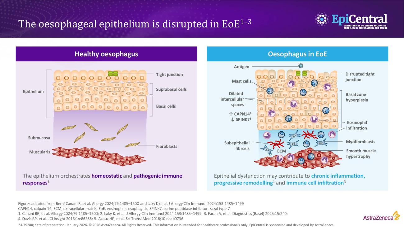
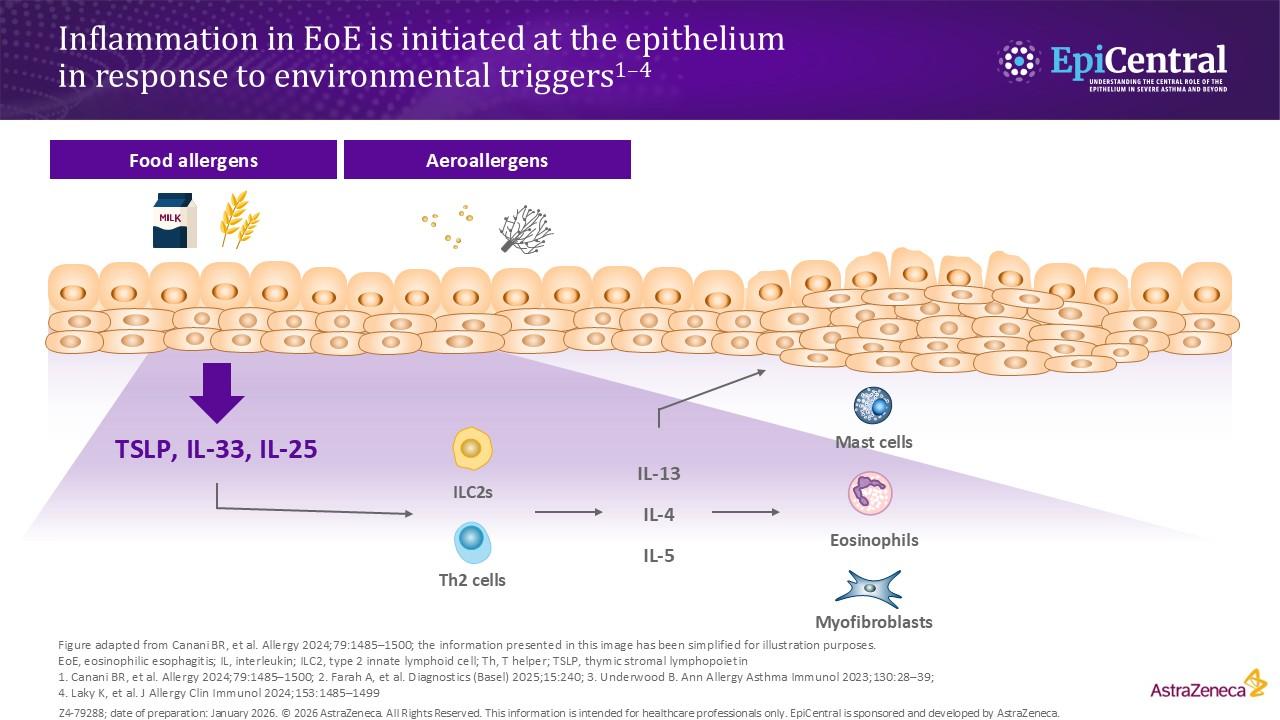
Ingested food allergens, including cow’s milk, wheat, soy and eggs, are known triggers of epithelial dysfunction and T2 inflammation in EoE, and are considered to be risk factors and drivers of the disease.4,8,14 A disrupted epithelial barrier permits allergen penetration and subsequent immune activation.8 Although food allergens are known triggers of EoE, the mechanisms behind allergen-specific immune activation are not fully understood and further research is needed. However, the removal of specific foods, either by dietary elimination or use of hypoallergenic formulas, can lead to disease remission.14

The involvement of aeroallergens, infectious disease and microbiome-altering factors in infancy (including antibiotic use during the first year of life, Caesarean delivery and pre-term birth, use of acid suppressants or a stay in a neonatal intensive care unit) have been also been implicated in several studies.14,15 Although these factors are being investigated for a possible role in EoE, they have not yet been defined as proven risk factors for EoE.15

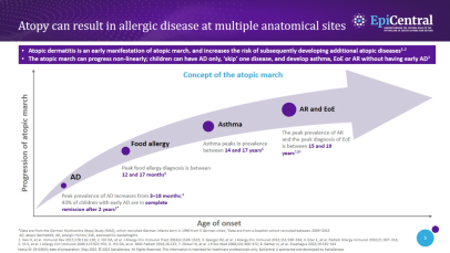
It has been established that EoE is over five-times more likely to occur in people with atopy, and the more atopic comorbidities a patient has, the more likely they are to have EoE.16,17 Much like EoE, atopic diseases, such as asthma, feature a disrupted epithelial barrier, which initiates and drives long-term T2 inflammation and leads to clinical symptoms.4,18 Previous research suggests that between 60% and 80% of patients have concomitant allergic conditions, including IgE-mediated food allergies, asthma, atopic dermatitis and allergic rhinitis;4 therefore, the more atopic comorbidities a patient has the more likely they are to have EoE.4,17,19,20 This highlights the importance of evaluating patient history when making a diagnosis.

The importance of the epithelium and its interaction with the environment in disease pathogenesis has also been seen in children with EoE. A recent study assessed for concurrent skin and esophageal dysfunction by evaluating ceramide levels in the skin of paediatric patients with EoE. Skin lipid composition of paediatric patients with EoE, but without atopic dermatitis (n=21), were compared with non-atopic dermatitis, non-EoE controls (n=17). It was found that the skin of patients with EoE have significant deficits in ceramide levels, particularly ultralong-chain fatty acid-containing ceramides, compared with the controls.21 This finding supports the suggestion that unified epithelial barrier dysfunction may underlie both skin and oesophageal atopic diseases.21

Want to learn more about role of epithelial cytokines in atopic diseases? Explore our downloadable resource on atopic march, developed in collaboration with Professor Marco Caminati, here

Learn more about epithelium-driven diseases, including asthma and chronic rhinosinusitis

Although EoE is believed to be a primarily environmentally driven disease, genetic factors, with familial associations, are also associated with the risk of developing EoE.12,22–24 Studies have found that adult first-degree relatives of patients with EoE are at risk for EoE, particularly those who are younger, male and have a history of atopic symptoms or diseases.17,23,24 The prevalence of EoE in first‑degree relatives ranges from 1.8% to 14.6%23,24 and the risk of developing EoE for siblings was 2.4%, increasing to 22% for dizygotic twins;23 these relatively low percentages also highlight the importance of environmental factors in EoE.23

Several studies, including candidate-gene identification and genome-wide association studies, have identified multiple genes, including TSLP and CAPN14, that may contribute to the development of EoE.12,22,25 The overexpression of these genes have been found to disrupt the oesophageal epithelial barrier and enhance immune-mediated T2 inflammation.12,22,25

Genomic analyses have identified polymorphisms in TSLP and TSLP receptor (TSLPR) associated with EoE.26–28 Single nucleotide polymorphisms residing in the TSLP gene were found to augment T2 responses, and play an important role in promoting T helper type 2 (Th2) cell differentiation, a potential immune cell contributing to oesophageal inflammation.22,25

CAPN14 encodes an interleukin (IL)-13 induced, oesophagus-specific proteolytic enzyme involved in epithelial homeostasis and repair, and may account for the tissue specificity of oesophageal disease in EoE.22,25

The overexpression of several other genes have been found to disrupt the oesophageal barrier and contribute to the development of EoE, including EMSY, STAT6, LRRC32, ANKRD27,CCL267, PDCD5, TGFB and HIF1α.7,8,22,25,29

Chronic oesophageal inflammation is associated with long-term complications in EoE

Over time, an inflammatory phenotype in EoE, primarily seen in children, may progress to a fibrostenotic or mixed inflammatory-fibrostenotic phenotype, typically seen in adults.30,31

Inflammatory phenotypes are associated with the early course of the disease and are commonly seen with children.30,31 Features of the inflammatory phenotype include: furrows, plaques, white exudates and a normal-presenting oesophagus with no evidence of fibrostenotic changes.30,31 Mixed and fibrostenotic phenotypes are associated with disease progression and the subsequent associated symptoms:4,30,32,33

  • Oesophageal fibrostenosis and reduced distensibility, requiring dilation
  • Food impaction requiring endoscopic bolus removal
  • Oesophageal perforation

Increasing fibrostenosis with age indicates that the natural history of EoE is a progression from an inflammatory to a fibrostenotic phenotype in the majority of patients.30 A retrospective Swiss study (N=200), found that 17% of patients with untreated EoE and 0–2 years of symptom duration had strictures, compared with 38% with >8–11 years of symptom duration and 71% with more than 20 years.34 Therefore, it may be hypothesised that treatment of chronic T2 inflammation could prevent disease progression.30 Understanding the pathogenesis of EoE and the early recognition of phenotypes may also aid physicians when making treatment decisions and improve outcome assessments.

Patients with EoE often experience years of diagnostic delay, which is associated with increased risks, including a 9% increased risk of strictures for every year of delay.30,31,35,36 Reasons for diagnostic delay include:

  • Patients using adaptive eating behaviours that can mask their symptoms4,37
  • Patients presenting with non-specific symptoms37
  • Lack of clinical suspicion or histological misinterpretation38
  • Lack of access to healthcare, issues with hidden costs such as travel to specialists or lacking necessary health insurance, when required39,40
  • Delayed endoscopy38

Pathophysiology: the central role of the epithelium in homeostasis and disease

The healthy oesophageal epithelium orchestrates homeostatic and pathogenic immune responses;6 intercellular junctional complexes (e.g., tight junctions) control paracellular transport and basal zone of epithelium occupies less than 15% of total epithelial thickness.6,7

In EoE the oesophageal epithelium is disrupted. This is characterised by:

  • Loss of structural proteins: E-cadherin, claudins and desmoglein-1, and an imbalance between increased protease expression (CAPN14) and decreased protease inhibitor expression (SPINK7) weakens tight junctions and adherens junctions, facilitating allergen penetration and immune activation8,41,42
  • Disrupted tight junctions: apical junction complex defects and dilated intercellular spaces are observed6,7
  • Basal zone hyperplasia: the basal cell layer occupies more than 15% of the total epithelial thickness43,44
  • Subepithelial fibrosis and smooth muscle hypertrophy can also be seen6,7

Image
Diagram illustrating how signalling pathways involved in inflammation in EoE are initiated in response to environmental triggers
Inflammation in EoE is initiated at the epithelium in response to environmental insults
Download resource

In EoE, the disrupted oesophageal epithelium interacts with environmental insults.6,8 Upon allergen exposure, epithelial cells release TSLP, IL-33 and IL-25, which activate Th2 cells (via dendritic cells) and type 2 innate lymphoid cells (ILC2s). These cells, in turn, secrete T2-polarising cytokines, including IL-4, IL-5 and IL-13, amplifying the inflammatory cascade, and contributing further to impaired epithelial barrier function.6,8,45 The chronic T2 inflammation may lead to fibrosis and remodelling, including strictures.7,8

Pathophysiology: the role of epithelial cytokines in EoE

Oesophageal biopsies from patients with EoE show significantly higher epithelial-derived cytokine expression than healthy controls.1,2,46 Variants in the genes encoding the alarmin TSLP are associated with increased risk of developing EoE, with TSLP expression found to be increased in patients with EoE.7 The overexpression of several critical genes, including genes encoding TSLP, may disrupt the oesophageal barrier and enhance immune-mediated inflammation.25

A study investigating the presence and distribution of eosinophil extracellular trap (EET) in oesophageal tissues from patients with EoE compared with healthy controls found EET formation was associated with defects of the epithelial barrier. Increased production of antimicrobial peptides and epithelial cytokines, known to drive Th2 inflammation and eosinophil activation, were also observed.2 When comparing active EoE with normal oesophageal samples, all three epithelial cytokines (TSLP, IL-33 and Il-25) showed increased expression.2 TSLP expression was found to correlate with the numbers of EET-positive eosinophils, suggesting a direct effect of TSLP on eosinophil functionality;2 however, IL-33 expression was not correlated with, and IL-25 expression was negatively correlated with, EET numbers.2

In a separate biopsy study, TSLP responsive-CD4+ T cells were shown to be increased in patients with EoE compared with non-EoE controls.47 TSLP responsive-CD4+ T cells (i.e. those expressing a receptor for TSLP) had augmented production of IL-4 or IL-13 compared with non-TSLP responsive-CD4+ T cells.47 In addition, TSLP was found to induce the expression of TSLPR, providing evidence of a feed-forward loop and a driver of T2 inflammation.47

Elevated IL-33 messenger RNA expression has also been observed in paediatric patients with confirmed EoE (n=9), when compared with paediatric patients without EoE (controls; n=5).48 Analysis of biopsy samples from patients with EoE detected that the IL‑33 receptor, ST2, has been detected on oesophageal-infiltrating eosinophils from patients with active EoE.49

Tumour necrosis factor-like ligand 1A (TL1A) is a newly discovered epithelial cytokine.50 Principally, TL1A has been found to synergise with IL-15 to induce several cytokines involved in the proinflammatory response,51 and has been implicated in inflammatory diseases of the gut;50 however, the role of TL1A in EoE remains poorly characterised.50

Download this slide deck to learn more about role of epithelial cytokines in patients with EoE

Evidence from EoE murine models has demonstrated that the epithelial cytokines (TSLP, IL-25 and IL-33) are elevated in EoE and play an important role in the symptomology and pathophysiology of EoE.52–54 EoE murine models have also indicated environmental triggers that may contribute to EoE pathogenesis, including, but not limited to, aeroallergens, food allergens and infectious diseases.7,55 Interestingly, a study by Doyle et al. also demonstrated that exposure to detergents, such as sodium dodecyl sulphate (SDS), found in common household products (e.g., toothpaste) decreases oesophageal barrier integrity, stimulates IL-33 production, and promotes epithelial hyperplasia and tissue eosinophilia in murine models (exposed to 0.5% SDS for 14 days).55 These animal models, therefore, support the important role of impaired epithelial health (i.e. epithelial barrier dysfunction, and subsequent immune cell recruitment and activation) in EoE.52–55

To find out more about epithelial cytokines in experimental models, click here

There is an unmet need for effective treatments for EoE

Based on current guidelines, the level of evidence to support the use of available therapies in patients with EoE is generally assessed to be low to moderate and there is a need for additional studies, including placebo-controlled studies, to assess the efficacy of therapies in EoE.3,4 There is currently no consensus on the recommended first-line therapy for EoE; however, there are several therapeutic options for patients with EoE:3,4

  • Swallowed topical corticosteroids (STCs), proton pump inhibitors (PPIs) and dietary elimination are all recommended treatments3,4
    • Feeding therapy can be used adjunctively in children with a food aversion or feeding dysfunction4
  • Biologics are currently considered to be a step-up therapy in difficult-to-treat patients or patients with multiple atopic conditions4
  • Oesophageal dilation is used as an adjunct to medical therapy, as a treatment for oesophageal strictures causing dysphagia4

More convenient dosing regimens and reduced cumulative side effects, currently associated with long-term treatment of existing medical therapies (such as those associated with corticosteroids), are required to improve patient adherence and perspectives.5 Although treatment with PPIs and STCs can be effective in some patients, response rates vary, with approximately two-thirds and one-third of patients, respectively, not achieving histological remission.5 Furthermore, wider issues, such as loss of response over time and lack of predictors of response, can all pose challenges with conventional pharmacological therapies.4,5,39,56

Dietary elimination is an effective, long-term option for patients with EoE. However, issues with response and adherence are reported.4,5 Studies have shown that half of patients do not maintain long-term diets and 30–65% of patients do not find dietary elimination effective (ranging across a one-food to six-food elimination diet).4 Repeated endoscopies, close monitoring and input from a dietician are required to support dietary elimination.4 A possible solution is to offer ‘diet holidays’, when patients can take a break from the diets for a set period of time, and bridge the break with medication.4

Additional research is needed to guide treatment decision-making when selecting first-line therapies for patients with different phenotypes.5 Shared decision-making with patients is key to ensuring optimum adherence and clinical outcomes.5

Despite recent advances in the diagnosis and management of EoE, challenges with the currently available treatments for EoE remain;5 future treatment should focus on where the disease initiates and, ultimately, restore the integrity of the epithelial barrier. Restoration through controlling chronic inflammation and preventing remodelling of the epithelium and progression of the disease are among the key considerations for future management of EoE.5

The content for this module was created with the support of Professor Seema Aceves, Professor Arjan Bredenoord and Professor Evan Dellon.

RELATED RESOURCES

Like this page? We have a host of other resources available in the ‘Scientific and Resource Library’ where you can find out more.

References

1. Travers J, et al. Sci Rep. 2017;7:17563; 2. Simon D, et al. Allergy. 2015;70:443–452; 3. Lucendo AJ, et al. United Eur Gastroenterol J. 2017;5:335–358; 4. Dellon ES, et al. Am J Gastroenterol. 2025;120:31–59; 5. Bredenoord AJ, et al. Am J Gastroenterol. 2022;117:1231–1241; 6. Berni Canani R, et al. Allergy. 2024;79:1485–1500; 7. Laky K, Frischmeyer-Guerrerio PA. J Allergy Clin Immunol. 2024;153:1485–1499; 8. Farah A, et al. Diagn Basel. 2025;15:240; 9. de Bortoli N, et al. Dig Liver Dis. 2024;56:951–963; 10. American Academy of Pediatrics. Diagnosis of eosinophilic esophagitis. 2023. Available from: https://www.aap.org/en/patient-care/eosinophilic-esophagitis/diagnosis-of-eosinophilic-esophagitis/?srsltid=AfmBOorPaMCbTZjQ_PagA339jwUq-5nLjl3g0RWIcrxEJtOSIyqgnMR_ (Accessed 22 January 2026); 11. Collins MH, et al. Esophagus. 2017;30:1–8; 12. Dhar A, et al. Gut. 2022;71:1459–1487; 13. Hahn JW, et al. Clin Gastroenterol Hepatol. 2023;21:3270–3284; 14. Dellon ES, Hirano I. Gastroenterology. 2018;154:319–332; 15. Jensen ET, Dellon ES. J Allergy Clin Immunol. 2018;142:32–40; 16. Weir AA, et al. Ann Allergy Asthma Immunol. 2025;134:362–364; 17. Hill DA, et al. J Allergy Clin Immunol Pr. 2018;6:1528–1533; 18. Akdis CA. Nat Rev Immunol. 2021;21:739–751; 19. Jaros J, et al. J Clin Aesthet Dermatol. 2025;18:15–20; 20. Hill DA, et al. J Allergy Clin Immunol Pr. 2017;5:369–375; 21. Goleva E, et al. J Allergy Clin Immunol. 2025;155:2075–2079; 22. O’Shea KM, et al. Gastroenterology. 2018;154:333–345; 23. Alexander ES, et al. J Allergy Clin Immunol. 2014;134:1084–1092; 24. Peterson K, et al. Clin Gastroenterol Hepatol. 2022;20:e957–e963; 25. Ryu S, et al. Int J Mol Sci. 2020;21:7253; 26. Rothenberg ME, et al. Nat Genet. 2010;42:289–291; 27. Sherrill JD, et al. J Allergy Clin Immunol. 2010;126:160–165; 28. Kottyan LC, et al. Nat Genet. 2014;46:895–900; 29. Masterson JC, et al. J Clin Invest. 2019;129:3224–3235; 30. Dellon ES, et al. Gastrointest Endosc. 2014;79:577–585; 31. Warners MJ, et al. Am J Gastroenterol. 2018;113:836–844; 32. Chen JW, et al. Endoscopy. 2016;48:794–801; 33. Runge TM, et al. J Clin Gastroenterol. 2017;51:805–813; 34. Schoepfer AM, et al. Gastroenterology. 2013;145:1230–1236; 35. Reed CC, et al. Clin Gastroenterol Hepatol. 2018;16:1667–1669; 36. Navarro P, et al. United Eur Gastroenterol J. 2022;10:507–517; 37. Muir AB, et al. Clin Exp Gastroenterol. 2019;12:391–399; 38. Kanakala V, et al. Eur J Gastroenterol Hepatol. 2010;22:848–855; 39. Dorrell R, et al. Gastroenterology. 2020;158(Suppl. 1):(Abstract S828); 40. Dellon ES. Ann Allergy Asthma Immunol. 2019;123:166–172; 41. Azouz NP, et al. Sci Transl Med. 2018;10:eaap9736; 42. Davis BP, et al. JCI Insight. 2016;1:e86355; 43. Collins MH. Gastrointest Endosc Clin N Am. 2008;18:59–71; viii–ix; 44. Collins MH. Dig Dis. 2014;32:68–73; 45. Underwood B, et al. Ann Allergy Asthma Immunol Off Publ Am Coll Allergy Asthma Immunol. 2023;130:28–39; 46. Chandramouleeswaran PM, et al. PLoS One. 2016;11:e0150968; 47. Rochman Y, et al. Sci Signal. 2023;16:eadg6360; 48. Judd LM, et al. Am J Physiol Gastrointest Liver Physiol. 2016;310:G13–G25; 49. Uchida AM, et al. Allergy. 2022;77:656–660; 50. Varricchi G, et al. J Allergy Clin Immunol. 2025;155:1420–1434; 51. Holmkvist P, et al. Mucosal Immunol. 2015;8:545–558; 52. Collison AM, et al. J Allergy Clin Immunol. 2015;136:971–982; 53. Dsilva A, et al. Allergy. 2025;doi 10.1111/all.16682 [Epub ahead of print]; 54. Masuda MY, et al. J Allergy Clin Immunol. 2024;153:1355–1368; 55. Doyle AD, et al. Allergy. 2023;78:192–201; 56. Haasnoot ML, et al. Am J Gastroenterol. 2022;117:1412–1418.