Achieving remission in airway diseases

Restoring epithelial health: can this lead to remission in patients with airway diseases?

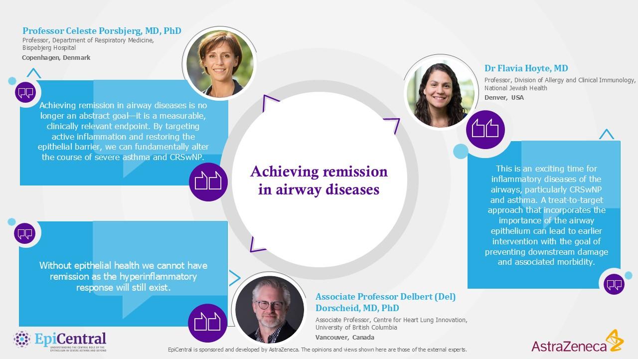
Remission should be a treatment goal in airway diseases

  • There is a lack of consensus around the definition of remission in asthma, allergic rhinitis and chronic rhinosinusitis with nasal polyps (CRSwNP), but proposed criteria include, among others, the absence of symptoms and a reduction in background medication use1–3
  • Remission while on biologics is possible. Approximately one in five patients with severe asthma treated with biologics achieve remission after 1 year4
  • Restoring epithelial health through repair of the epithelial barrier is an important step on the road to remission.5–8
  • Mechanisms of epithelial repair, including increasing tight junction expression and normalising ciliary beat frequency, contribute to improvements in clinical outcomes5,7,9–13
  • The goal of achieving remission should be to reduce disease activity to promote repair of damaged airways.1,3,7,14
  • Using targeted therapies, the epithelial barrier can be restored, preventing the loss of lung function, decreasing exacerbations, and has the potential to reduce mortality1,4,15–17

1. Caminati M, et al. Curr Allergy Asthma Rep. 2024; 24(1): 11–23; 2. Carpaij OA, et al. Pharmacol Ther. 2019; 201: 8-24; 3. Chan Y, et al. J Otolaryngol Head Neck Surg. 2023; 52(1): 50; 4. Perez-de-Llano L, et al. Am J Respir Crit Care Med. 2024;210(7):869-880; 5. Russell RJ, et al. Eur Respir J. 2024;63(4): 2301397; 6. Davies DE. Proc Am Thorac Soc. 2009;6(8):678-82; 7. Brightling CE, et al. Eur Respir Rev. 2024;33(174):240221; 8. Heijink IH, et al. Allergy. 2020;75(8):1902-17; 9. Kempeneers C, et al. ERJ Open Res. 2023;9(5):00220-2023; 10. Noureddine N, et al. J Asthma Allergy. 2022;15:487-504; 11. Ghezzi M, et al. Children (Basel). 2021;8(12):1167; 12. Georas SN, Rezaee F. J Allergy Clin Immunol. 2014;134(3):509-20; 13. Tilley AE, et al. Annu Rev Physiol. 2015;77:379-406; 14. Thomas D, et al. Eur Respir J. 2022;60(5):2102583; 15. Pelaia C, et al. J Clin Med. 2023;12(10):3371; 16. Maspero J, et al. ERJ Open Res. 2022;8(3):00576-2021; 17. Mailhot-Larouche S, et al. Ann Allergy Asthma Immunol. 2024:S1081-1206(24)01559-X.

Image
KEE quotes
Expert quotes on remission
Download resource

Remission as a clinical endpoint in upper and lower airway disease

Airway diseases, such as asthma, allergic rhinitis and chronic rhinosinusitis with nasal polyps (CRSwNP), are conditions characterised by chronic inflammation that cause long-term damage and potentially irreversible airway remodelling.1-3 Addressing the immune response and controlling disease activity to reduce inflammation is a key step to achieving remission and requires management of immune dysregulation and restoration of epithelial health.3-8 There has been considerable evolution in the management of airway diseases, particularly over the past 30 years, leading to the development of biologics that target specific inflammatory pathways and can be used by patients to achieve remission.1,9-13

Adopting a treat-to-target approach is currently being discussed; a treatment strategy wherein the clinician treats the patient proactively enough to reach and maintain explicitly specified and sequentially measured goals. In this scenario remission would be the treatment target, with primary goals to eliminate symptoms and exacerbation risk, prevent airway remodelling and normalise lung function. Achieving these targets would lead to better patient outcomes.14,15

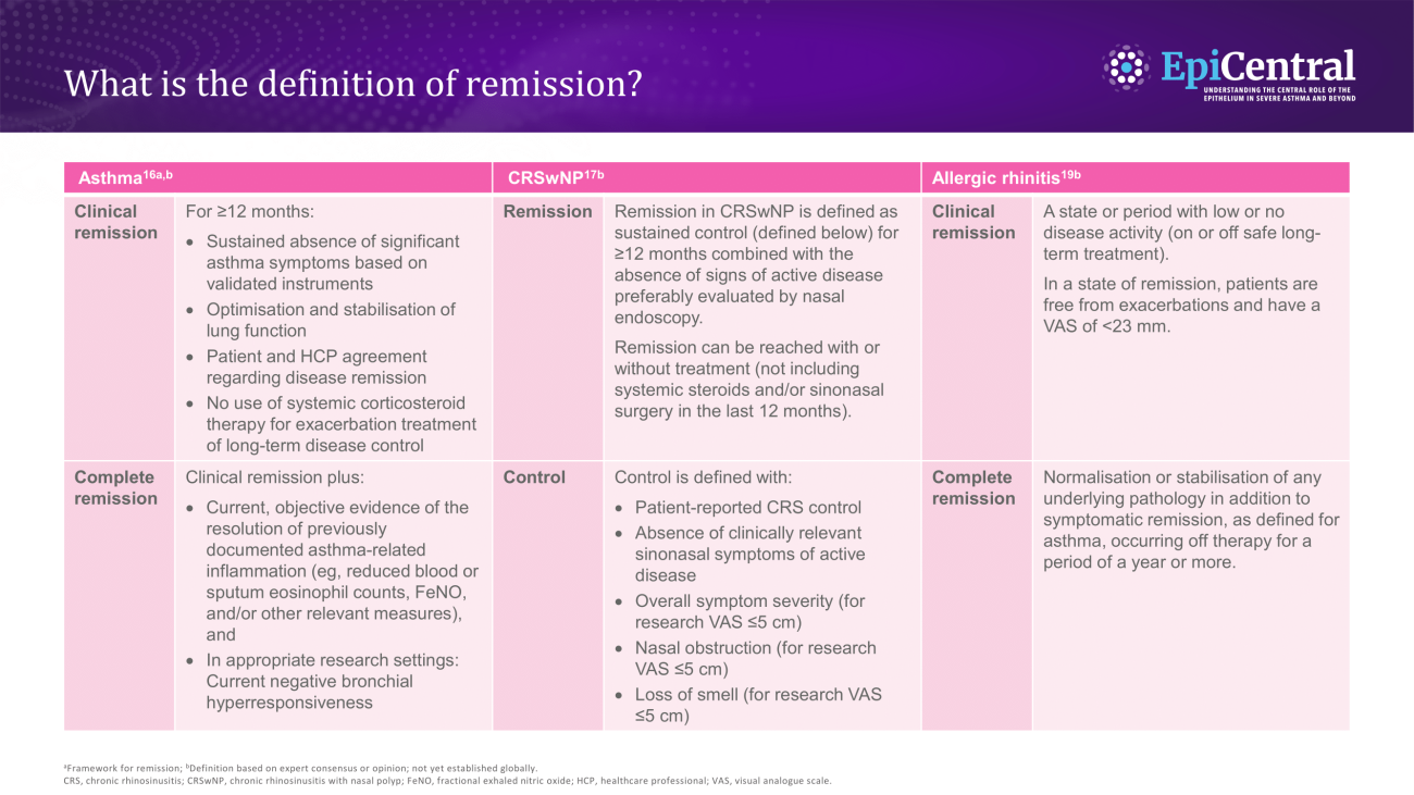
The proposed definition of clinical remission in asthma, CRSwNP and allergic rhinitis has evolved over time,16,17 with a definition previously focused on spontaneous or off-treatment remission, but has now evolved to focus on remission in patients who remain on treatment.3,18 Multiple criteria for remission in airway diseases have been proposed.16,17 These criteria have been developed based on definitions of remission in rheumatoid arthritis, ulcerative colitis, Crohn’s disease and systemic lupus erythematosus, where the remission is a recognised clinical endpoint which has supported treatment advances and improved patient outcomes.16 Broadly speaking, proposed definitions of remission in asthma include four components: no exacerbations, symptom control, no need for maintenance OCS (mOCS), and optimisation and stabilisation of lung function within a defined window.16 In CRSwNP, remission is suggested to be defined as sustained control (patient-reported chronic rhinosinusitis [CRS] control, absence of clinically relevant sinonasal symptoms of active disease, lack of nasal obstruction, no loss of smell) for ≥12 months.17 The definition of remission in allergic rhinitis is more sparsely explored, but a proposed definition includes a state or period with low or no disease activity, preferably with a normal nasal examination.19

There is limited evidence on the rates of on-treatment remission in airway diseases based on these definitions.3,16,17,20 Following the more widespread adoption of biologics, a longitudinal cohort study has shown that one in five patients with severe asthma achieved four-domain remission16 within one year of biologic initiation.13 CRSwNP studies of endoscopic sinus surgery with continued medical therapy reported remission rates of approximately 50%,20 while in an allergic rhinitis longitudinal study, 32% of self-reported patients no longer reported the condition ~10 years later, which investigators considered as being in remission.21

Barrier dysfunction: Histological changes and functional abnormalities of the epithelial barrier resulting from repeated injury, repair and regeneration of the epithelium22
Disease activity: Underlying manifestations of disease, as determined by symptoms, exacerbation frequency and response to treatment23,24
Epithelial health: Descriptor of epithelial integrity and function; can be altered by damage or repair to the epithelium25,26
Epithelial repair: Restoration of a damaged epithelium through interactions between immune cells and the epithelium to maintain epithelial integrity27
Remission: A state or period with low to no disease activity that can be spontaneous or a result of therapy16

The critical role of the epithelium in the immune response in upper and lower airway disease

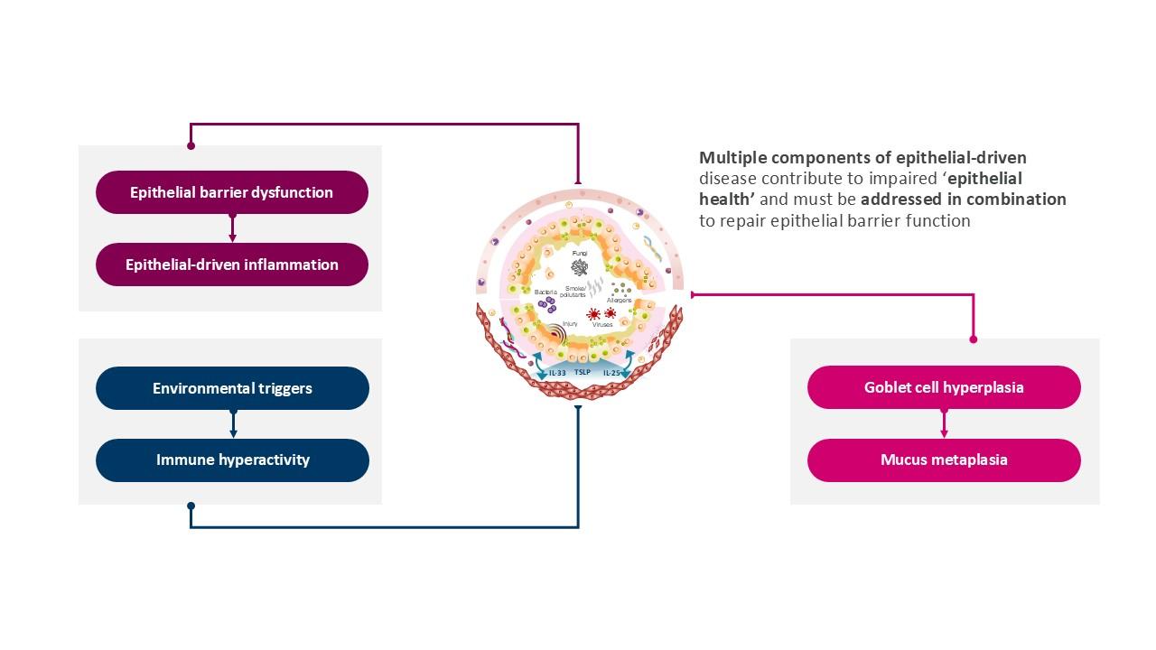
The epithelium does not just act as a physical barrier but also initiates and controls the inflammatory response to injury and has a homeostatic function.28,29 A dysfunctional and activated epithelium is a critical component of airway diseases like severe uncontrolled asthma and CRSwNP.5,27,30 In people with severe asthma, the epithelium initiates and amplifies an inflammatory feedback loop, characterised by an excessive production of proinflammatory cytokines; a critical component of disease activity.4,5,7 Addressing the immune response and controlling disease activity to reduce inflammation is a key step to achieving remission and requires management of immune dysregulation and restoration of epithelial health.3-7

In patients with airway diseases, the epithelium cannot process external signals and generates an excessive immune response event to usual triggers, which leads to a gradual loss of lung function in patients with asthma.1,7 The epithelium drives the exaggerated ‘hyper-response’ through the release of epithelial cytokines that is characteristic of the epithelial barrier disruption seen in asthma and CRSwNP.7,30,31 Epithelial barrier disruption is sustained and reinforced through positive feedback. Consequently, the excessive release of epithelial inflammatory cytokines feeds back on itself, which contributes to epithelial barrier dysfunction.5,7 One example is in atopic individuals wherein allergen inhalation leads to mast cell degranulation, and mast cell mediators increase epithelial permeability, which predisposes these patients to further allergic sensitisation and increases the interaction of the epithelium with environmental triggers.5,32

The downstream effects of the epithelial immune hyper-response are numerous and expansive; establishing the epithelium as a key coordinator of airway diseases, and therefore an important target.7,30-32

Compromised epithelial health leads to barrier dysfunction, chronic epithelial-driven inflammation and disease7,25,33-36

Image
Epithelial dysfunction figure
IL, interleukin; TSLP, thymic stromal lymphopoietin.
Download resource

Mucus hypersecretion
In patients with asthma, mucus hypersecretion is driven by increased IL-13; a downstream effect of the inflammatory cascade triggered by the release of epithelial cytokines.37 Increased IL-13 is associated with airway hyperresponsiveness (AHR) to methacholine. In vitro studies of epithelial cells show that IL-13 significantly increases expression of MUC5AC, an integral component of mucus.38 Coupled with goblet cell hyperplasia, this can lead to the development of mucus plugs.7,38 Mucus plugging is associated with eosinophilia and increased type 2 (T2) cytokine production, and correlates with the severity of airflow obstruction and lung function decline.7


Inflammation
Epithelial cytokines drive T2 inflammation; a response to injury driven by the innate and adaptive immune system through the production of T2 cytokines by type 2 innate lymphoid cells and type 2 T-helper cells, respectively.2 T2 inflammation, along with exacerbations and AHR, correlate with lung function decline.7,39-41 Blood eosinophils and fractional exhaled nitric oxide (FeNO), markers of T2 inflammation, are associated with accelerated lung function decline.39 This correlation has been seen across individuals with chronic airway disease, but is more pronounced in people with asthma.39 In keeping with the united airways theory, co-morbid asthma and CRSwNP is associated with significantly increased T2 inflammation as demonstrated by increased blood and sputum eosinophils, and IgE.42,43


Exacerbations
Exacerbations are markers of disease activity that are driven by the T2 inflammation initiated by the epithelium.7,44 Epithelial cytokines likely contribute to exacerbation as blocking certain cytokines reduces exacerbation rates.7 Exacerbations lead to airway remodelling and reduced lung function.40,45 Patients on high doses of ICS experience the fastest decline in lung function, indicating that the pathways driving exacerbations are not fully suppressed by ICS.40 AHR has been shown to be independently associated with accelerated decline in lung function irrespective of age, gender, smoking status, respiratory symptoms and baseline FEV1 (forced expiratory volume in 1 second).41 AHR also has a negative effect on lung function development from childhood to adulthood.46 Patients with airway disease may present with abnormal basal cell differentiation, indicating an impaired repair mechanism.5,32 Together with reduced ciliated cells, goblet cell hyperplasia and increased epithelial mesenchymal transition, this can contribute to impaired mucociliary clearance and airway remodelling.32


Airway remodelling
Epithelial cytokines, IL-25, IL-33 and TSLP, play diverse and often overlapping roles in airway remodelling, including the proliferation of lung fibroblasts, expression of collagen and fibronectin and smooth muscle wound repair.37,47-49 Together with other T2 cytokines and factors, such as the epidermal growth factor receptor, these cytokines drive the repair of the epithelium through normal and abnormal processes.32 Various triggers at the level of the epithelium initiate excessive release of IL-25, IL-33 and TSLP. These cytokines, termed alarmins, are upstream drivers of disease that influence multiple aspects of the inflammatory cascade, leading to airway AHR and irreversible structural changes.7,37 They are integral to the pathogenesis of multiple airway diseases and multiple disease phenotypes.1,2,37

To learn more about the role of the epithelium in asthma, please click here

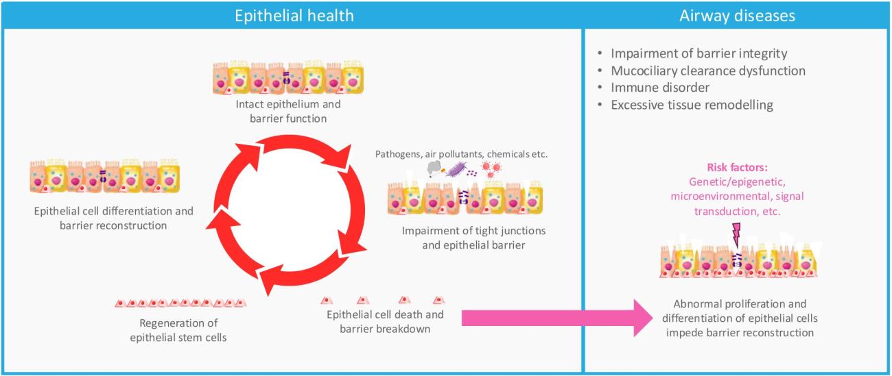
Epithelial health and dysfunction50

Image
Epithelial health and dysfunction
Download resource
Restoring epithelial health: evidence for epithelial repair?

To improve symptoms, we need to restore epithelial health. Epithelial health is dependent on many contributing factors, including healthy interaction of the epithelium and the immune system, and intact airway physiology and structure.32,51 In patients with asthma, epithelial health is diminished, leading to impaired innate defence, persistent T2 inflammation, barrier disruption and tissue remodelling.7 Restoring epithelial barrier function through epithelial repair could move patients closer to a healthier epithelium, where the immune response to environmental exposures and insults is proportional and sufficient, improving their chances of achieving remission.7,25,35,36

To achieve remission, the epithelium must be repaired and epithelial health restored.25,52 In in vitro studies, barrier function was restored following treatment with epidermal growth factor, indicating that tight junction expression can be increased and that defective airway barrier function in asthma is potentially reversible.52 Repairing airway epithelial cells likely forms a key part of restoring epithelial health.7,53 Ciliary beat frequency needs to be normalised and mucus-producing cells reduced to increase mucociliary clearance and prevent the development of mucus plugs and airway occlusion.7,54,55 Reversing airway remodelling, which is partially characterised by thickening of the airway epithelium, can relieve asthma symptoms and prevent disease progression, but it is acknowledged that reversing airway remodelling is difficult.56 Corticosteroids have been shown to not sufficiently address this pathophysiology and may even promote it.57 Reducing epithelial-mesenchymal transition, as triggered by epithelial injury, may be a mechanism to decrease airway remodelling.36,58,59

Improvements in clinical outcomes are a consequence of epithelial restoration and reduced epithelial barrier dysfunction.16,17,57,60,61 Restoring barrier function reduces interaction of triggers with the epithelium, which reduces inflammation; a driver of many symptoms in asthma and CRSwNP.61 T2 inflammatory markers positively correlate with symptoms in people with asthma and CRSwNP.39 In some studies in patients in remission, these inflammatory markers have been found to be expressed at lower levels suggesting that the epithelial barrier has been restored.62 For example, in asthma, reducing chronic T2 inflammation decreases AHR, which leads to fewer exacerbations.7,63 Patients with lower inflammatory marker expression also have fewer symptoms, which supports the idea that epithelial damage is associated with increased symptom burden, including reduced lung function in asthma and loss of smell, nasal obstruction and nasal discharge in CRSwNP.1,64

In asthma, AHR and airway remodelling are a consequence of damage and activation of the epithelium that leads to inflammatory signalling – preventing, reducing or repairing this damage could improve symptoms.1,7 Epithelial barrier damage and dysfunction are drivers of the pathophysiology of asthma and CRS, and restoration of epithelial structure and function has the downstream effect of helping patients achieve the proposed criteria of remission.1,7,16,17

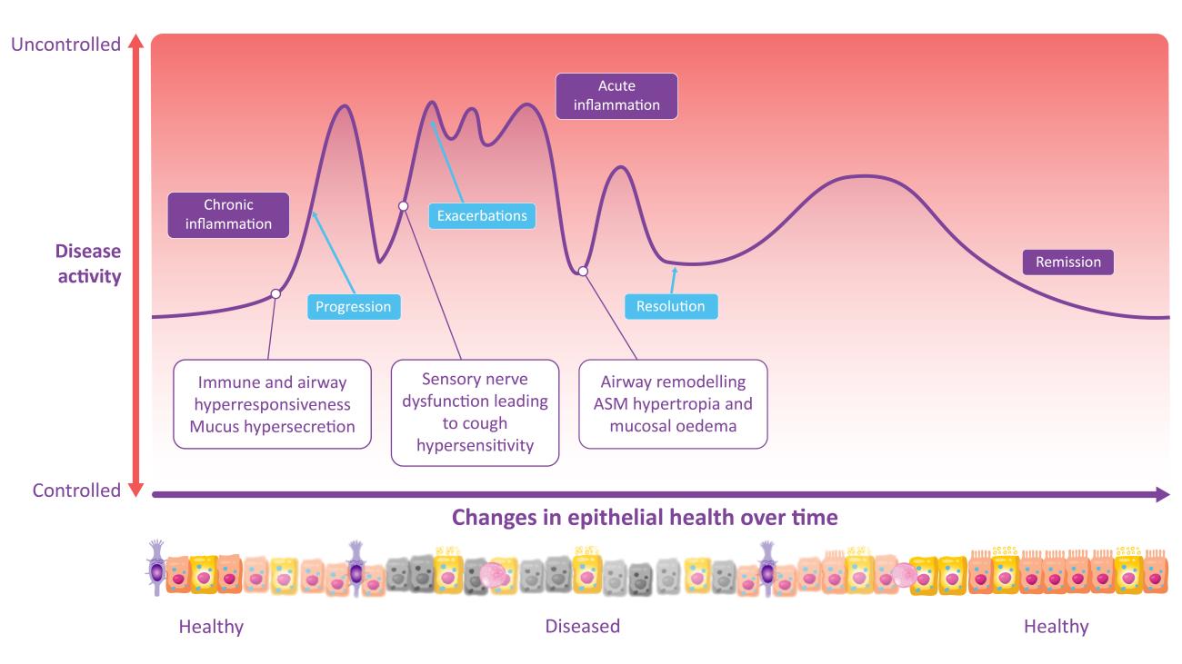
Relationship between disease activity, changes in epithelial health and clinical presentation of asthma32,33,65-67

Image
Effect of intervention on the clinical presentation of remission in asthma.
ASM, airway smooth muscle.
Download resource

Disease activity and damage – a dual approach to clinical remission

In previous discussions of remission, the ‘type’ of remission has not been consistent, nor has the underlying active disease manifestation always been considered.3,13 A dual approach to remission is now being considered, taking into account disease activity and damage.7,14,23,68,69 Disease activity describes the exaggerated response of the immune system triggered by the epithelium.7,30,31 Long-term damage includes remodelling and impaired epithelial cell function, and can lead to loss of lung function, development and worsening of comorbidities, and mortality.2,7,70,71

Reducing epithelial inflammation, through targeting inflammatory mediators before asthma severity escalates, could provide an opportunity to prevent epithelial damage and achieve remission.7,13,14,57 Early intervention using appropriate treatment may prevent the accumulation of irreversible damage and halt disease progression, making remission more likely.3,13,14,57 Controlling disease activity through reducing inflammation likely improves patients’ chance of remission particularly in the case of reducing the progression of airway remodelling.3,14,20,25

Remission has a positive impact on patient outcomes and should be the clinical goal in severe asthma, CRSwNP and allergic rhinitis.3,14 Remission is associated with improved quality of life through improved symptoms, fewer exacerbations, a reduced need for OCS and improved lung function.3,16,72 All of these outcomes are associated with a reduced healthcare resource utilisation.16,73,74

Next steps to achieving remission

The epithelium is a key driver of inflammatory pathways and therefore is an important target in treating airway diseases.1,2,7 Remission in airways diseases is a viable target. In one cohort study, clinical remission was achieved in ~20% of patients with asthma on biologics, while some clinical trials have reported rates that are even higher, but these rates vary between trials.13,71 The same cohort study suggested benefits of earlier intervention as chances of remission increased with shorter durations of asthma.13

Further research into remission in patients with airway diseases is needed, including:

  • What factors outside of asthma duration improves a patient’s chance of achieving remission?
  • What factors increase the risk of severe asthma and can help identify which patients require early intervention?
  • Can we identify better biomarkers for upper and lower airway disease that normalise in patients who achieve remission?
  • Is airway remodelling reversible in patients with asthma, and is this reversal required in some patients to induce remission?
  • How can the rates of remission be increased in patients with airway diseases?

RELATED RESOURCES

Like this page? We have a host of other resources available in the ‘Scientific and Resource Library’ where you can find out more.

References

1. Pelaia C, et al. J Clin Med. 2023;12(10):3371; 2. Maspero J, et al. ERJ Open Res. 2022;8(3):00576-2021; 3. Thomas D, et al. Eur Respir J. 2022;60(5):2102583; 4. Holgate ST. Allergol Int. 2008;57(1):1-10; 5. Hellings PW, Steelant B. J Allergy Clin Immunol. 2020;145(6):1499-509; 6. Chabra R, Gupta M. Treasure Island (FL): StatPearls Publishing; 2025. Available from: https://www.ncbi.nlm.nih.gov/books/NBK526018/ (Accessed 1 May 2025); 7. Russell RJ, et al. Eur Respir J. 2024;63(4):2301397; 8. Murphy RC, et al. J Allergy Clin Immunol Pract. 2021;9(7):2588-97; 9. Crompton G. Prim Care Respir J. 2006;15(6):326-31; 10. Tanaka A. J Gen Fam Med. 2015;16(3):158-69; 11. Pavord ID, et al. Lancet. 1999;353(9171):2213-4; 12. Hellings PW, et al. Allergy. 2024;79(5):1123-33; 13. Perez-de-Llano L, et al. Am J Respir Crit Care Med. 2024;210(7):869-80; 14. Caminati M, et al. Curr Allergy Asthma Rep. 2024;24(1):11-23; 15. Nannini LJ. J Asthma. 2020;57(6):687-90; 16. Menzies-Gow A, et al. J Allergy Clin Immunol. 2020;145(3):757-65; 17. Fokkens WJ, et al. Rhinology. 2024;62(3):287-98; 18. Lommatzsch M. Curr Opin Pulm Med. 2024;30(3):325-9; 19. Scadding GK, et al. Front Allergy. 2025;5:1531788; 20. Chan Y, et al. J Otolaryngol Head Neck Surg. 2023;52(1):50; 21. Heldin J, et al. J Asthma Allergy. 2022;15:1569-78; 22. Gon Y, Hashimoto S. Allergol Int. 2018;67(1):12-7; 23. Greenberg S, et al. J Allergy Clin Immunol. 2012;130(5):1071-7.e10; 24. Smith NMJ, et al. BMJ Open Respir Res. 2020;7(1); 25. Brightling CE, et al. Eur Respir Rev. 2024;33(174):240221; 26. Reynolds SD, et al. Proc Am Thorac Soc. 2012;9(2):27-37; 27. Inoue H, et al. J Clin Med. 2020;9(11); 28. Tam A, et al. Ther Adv Respir Dis. 2011;5(4):255-73; 29. Hewitt RJ, Lloyd CM. Nat Rev Immunol. 2021;21(6):347-62; 30. Kohanski MA, et al. J Allergy Clin Immunol. 2021;148(5):1161-4; 31. Kato A, et al. J Allergy Clin Immunol. 2022;149(5):1491-503; 32. Raby KL, et al. Front Immunol. 2023;14:1201658; 33. Porsbjerg C, et al. Lancet. 2023;401(10379):858-73; 34. Asthma CoEiS. 2018. Available from: https://toolkit.severeasthma.org.au/severe-asthma/pathophysiology/ (Accessed 1 May 2025); 35. Heijink IH, et al. Allergy. 2020;75(8):1902-17; 36. Davies DE. Proc Am Thorac Soc. 2009;6(8):678-82; 37. Gauvreau GM, et al. Expert Opin Ther Targets. 2020;24(8):777-92; 38. Bonser LR, Erle DJ. J Clin Med. 2017;6(12):112; 39. Çolak Y, et al. Thorax. 2024;79(4):349-58; 40. Soremekun S, et al. Thorax. 2023;78(7):643-52; 41. Kanemitsu Y, et al. Allergol Int. 2014;63(2):181-8; 42. Wang M, et al. Clin Transl Allergy. 2020;10:26; 43. Bilodeau L, et al. Rhinology. 2010;48(4):420-5; 44. Federico MJ, et al. J Allergy Clin Immunol. 2007;119(1):50-6; 45. Bai TR, et al. Eur Respir J. 2007;30(3):452-6; 46. Harmsen L, et al. Respir Med. 2014;108(5):752-7; 47. Guo Z, et al. J Asthma. 2014;51(8):863-9; 48. Kaur D, et al. Allergy. 2015;70(5):556-67; 49. Xu X, et al. Exp Biol Med (Maywood). 2019;244(9):770-80; 50. Huang ZQ, et al. Allergy. 2024;79(5):1146-65; 51. Fehrenbach H, et al. Cell Tissue Res. 2017;367(3):551-69; 52. Georas SN, Rezaee F. J Allergy Clin Immunol. 2014;134(3):509-20; 53. López-Posadas R, et al. Front Cell Dev Biol. 2024;12:1258859; 54. Tilley AE, et al. Annu Rev Physiol. 2015;77:379-406; 55. Kempeneers C, et al. ERJ Open Res. 2023;9(5):00220-2023; 56. Huang Y, Qiu C. Ann Transl Med. 2022;10(18):1023; 57. Varricchi G, et al. Eur Respir J. 2024;63(4):2301619; 58. Ha JG, Cho HJ. Int J Mol Sci. 2023;24(18):14229; 59. Sun Z, et al. Front Immunol. 2020;11:1598; 60. Noureddine N, et al. J Asthma Allergy. 2022;15:487-504; 61. Ghezzi M, et al. Children (Basel). 2021;8(12):1167; 62. Carpaij OA, et al. Pharmacol Ther. 2019;201:8-24; 63. Chapman DG, Irvin CG. Clin Exp Allergy. 2015;45(4):706-19; 64. AlBloushi S, Al-Ahmad M. Front Immunol. 2024;15:1285598; 65. Jesenak M, et al. Chest. 2024; 66. Cohn L. J Allergy Clin Immunol. 2022;150(1):59-61; 67. Porsbjerg C, et al. Remission and response in asthma and chronic obstructive pulmonary disease [Symposium: Session ID 271]. ERS, 7–11 September 2024, Vienna, Austria; 68. Al Ghobain MO, et al. Cureus. 2023;15(2):e35289; 69. Volbeda F, et al. Thorax. 2013;68(1):19-24; 70. O'Byrne P, et al. Eur Respir J. 2019;54(1):1900491; 71. Mailhot-Larouche S, et al. Ann Allergy Asthma Immunol. 2024;S1081-1206(24):01559-X; 72. Busse WW, et al. J Allergy Clin Immunol Pract. 2024;12(4):894-903; 73. Wisnivesky J, et al. J Asthma. 2023;60(6):1072-9; 74. Price D, et al. Eur Respir Rev. 2020;29(155):190151.