The importance of the epithelium and epithelial cytokines in uniting upper and lower airway diseases

Epithelial dysregulation is implicated in both upper and lower airway diseases.1,2

In epithelial-driven disease, multiple environmental insults cause release of epithelial cytokines (TSLP, IL-33 and IL-25),1,2 which results in the activation of innate and adaptive immune responses and the initiation of multiple inflammatory pathways. Epithelial barrier disruption is central to the pathogenesis of inflammatory conditions throughout the airway,3–5 and the frequent co-occurrence of upper and lower airway inflammatory disease has led to the ‘United airways disease’ concept.6–9

  • The upper and lower airways are linked anatomically, histologically and immunologically6,7
  • Epithelial barrier dysfunction and epithelial cytokines are linked to chronic inflammatory diseases of multiple organ systems, including lower airway diseases (eg asthma) and upper airway diseases (eg chronic rhinosinusitis and allergic rhinitis)6,10–12
  • Upper airway diseases have a negative effect on patients socially and psychologically, severely impacting their quality of life13,14
  • Chronic rhinosinusitis is a heterogeneous inflammatory condition that is characterised by mucosal inflammation of the nasal passage,15,16 which can be divided into two phenotypes based on the presence or absence of nasal polyps16–18
  • Rhinitis is inflammation of the mucous membrane lining in the nasal passages; it can be classified as allergic or non-allergic rhinitis13,19

Understanding the united pathology of upper and lower airway diseases suggests that a united approach to disease management may be needed to achieve global disease control for patients.9,20

1. Roan F, et al. J Clin Invest. 2019;129:1441–1451; 2. Mitchell PD, O’Byrne PM. Chest. 2017;151:1338–1344; 3. Hellings PW, Steelant B. J Allergy Clin Immunol. 2020;145:1499–1509; 4. Russell RJ, et al. Eur Respir J. 2024;63:2301397; 5. Ha J-G, Cho H-J. Int J Mol Sci. 2023;24:14229; 6. Fokkens W, Reitsma S. Otolaryngol Clin North Am. 2023;56:1–10; 7. Jakwerth CA, et al. Cells. 2022;11:1387; 8. Yii AC, et al. Allergy. 2018;73:1964–1978; 9. Kicic A, et al. J Allergy Clin Immunol. 2020;145:1562–1573; 10. Heijink IH, et al. Clin Exp Allergy. 2014;44:620–630.11. Bousquet J, et al. Nat Rev Dis Primers. 2020;6:95; 12. Bartemes KR, Kita H. Clin Immunol. 2012;143:222–235; 13. Dykewicz MS, et al. J Allergy Clin Immunol. 2020;146:721–767; 14. Bachert C, et al. J Asthma Allergy. 2021;14:127–134; 15. Lee S, Lane AP. Curr Infect Dis Rep. 2011;13:159–168; 16. Brzost J, et al. Diagnostics (Basel). 2022;12:2301; 17. Orlandi RR, et al. Int Forum Allergy Rhinol. 2016;6(Suppl. 1):S3–S21; 18. Fokkens WJ, et al. Rhinology. 2020;58(Suppl. S29):1–464; 19. Beard S. Prim Care. 2014;41:33–46; 20. Licari A, et al. Front Pediatr. 2017;5:44

Image
Expert Quotes - Dr Laidlaw and Professor Heffler

Insights from our EpiCollaborators

Download resource

Introduction to epithelial-driven diseases

In epithelial-driven disease, multiple environmental insults cause release of epithelial cytokines (thymic stromal lymphopoietin [TSLP], interleukin [IL]-33 and IL-25),1,2 which results in the activation of innate and adaptive immune responses and the initiation of multiple inflammatory pathways.1 Inflammation driven by epithelial cytokines can further disrupt the epithelial barrier.3 As such, epithelial barrier dysfunction is linked to chronic inflammatory diseases of multiple organ systems, including lower airway diseases (eg asthma and COPD) and upper airway diseases (eg chronic rhinosinusitis and allergic rhinitis).4,5

In the airway, internal and external insults cause epithelial barrier dysfunction via reduced tight-junction function,3 increased permeability,3 plasma leakage,6,7 goblet cell hyperplasia and/or increased mucus production.3,7 The active airway epithelium, in response to insults, also causes inflammation through the release of the epithelial cytokines.3

Download this slide deck to learn more about the role of the epithelium in airway disease.

Introduction to the united airways disease concept

The upper and lower airways are linked anatomically, histologically and immunologically.4,8 Pathological processes which occur in one part of the airway may affect the other; thus, although upper and lower airway diseases can present with different symptoms, the underlying immunological response is often similar.4,9 The frequent co-occurrence of upper and lower airway inflammatory diseases and the immunological links between the two parts of the airways is known as ‘United airways disease’.4,8–10

Upper airway diseases and the role of the epithelium

The nasal airway epithelium is the gateway to the respiratory system, acting as the first point of contact for viruses, pollutants, allergens and other airborne environmental triggers.11,12 Here, inhaled air is warmed, humidified and filtered, with larger particles trapped by hairs in the nose.4,11,13,14 Smaller particles invade deeper into the airway, where they are captured in secreted mucus and expelled by the action of ciliated cells, in a process known as mucociliary clearance.11,13,14 In addition to the physical barrier provided by the nasal mucosa, in healthy individuals, epithelial cells throughout the airways also initiate an immune response which removes invaders from the respiratory system.8,15,16

Just as epithelial dysregulation is implicated in the pathogenesis of asthma,5,16 disruption of the nasal epithelial barrier contributes to upper airway diseases (e.g., allergic rhinitis and chronic rhinosinusitis) through increased permeability and infiltration by external stimuli.4,12 Interaction with external stimuli leads to mucosal inflammation in the upper airways, resulting in the release of cytokines and inflammatory mediators.4,10,14 Inflammation is associated with upper airway remodelling, for example fibrosis, basement membrane thickening, goblet cell hyperplasia, polyp formation, osteitis and angiogenesis.14,17 Epithelial cytokines (TSLP, IL-33 and IL-25), released by the epithelium in response to triggers, have been implicated in the pathogenesis of upper airway diseases, such as chronic rhinosinusitis and allergic rhinitis.4,18

Broadly, the airway epithelium consists of a continuous, impermeable sheet of cells, held together with tight junctions and adhesion proteins, which sits on the basement membrane.11–13,19,20 In both the upper and lower airways, ciliated and secretory cells facilitate clearance of mucus and airway debris.11,13,14,20 In the upper airways, about 20% of the epithelium is composed of goblet cells,11 which produce mucus with antioxidant, antiprotease and antimicrobial properties.19 Moving into the lower airways and bronchioles, there are fewer goblet cells;11,19 instead, club cells secrete products that have anti-inflammatory and immunosuppressive capacity,11,20 which helps to maintain homeostasis.20

At the end of the bronchial tree, Type 1 alveolar cells, which facilitate gas exchange, and Type 2 alveolar cells, which secrete surfactants, form the alveoli.11 A key factor which differentiates the lower airway epithelium from the upper is the presence of smooth muscle, which drives bronchoconstriction.4,21
Image
UAD epithelial structure infographic

Chronic rhinosinusitis

Chronic rhinosinusitis (CRS) is characterised by chronic sinonasal inflammation and divided into two main phenotypes: CRS with nasal polyps (CRSwNP) and CRS without nasal polyps (CRSsNP).22 Estimates of CRS prevalence range from less than 5% of the population to more than 10%.23 The condition has a significant psychological and social burden, but patients perceive an underestimation of disease burden by healthcare professionals (HCPs).24

Epithelial dysfunction is an important characteristic of CRS and impedes the ability of the epithelium to act as a physical and immune barrier against the external environment.25 Elevated expression of epithelial cytokines has been demonstrated in the nasal epithelium of patients with CRSwNP,26 and levels of thymic stromal lymphopoietin (TSLP), the TSLP receptor (TSLPR) and the interleukin (IL)-33 receptor (ST2L) have been shown to correlate with markers of disease severity in eosinophilic CRSwNP.27

Click here to learn more about the central role of the epithelium in CRS

CRSwNP

In patients with a T2 endotype, epithelial disruption leads to a cycle of T2 inflammation.29 The defective epithelial barrier is more easily permeable, and epithelial cytokines (TSLP, IL-33 and IL-25) are released in response to external triggers acting on the epithelium.15,29 All three epithelial cytokines have been shown to be overexpressed in patients with CRSwNP.30–32

Image
CRSwNP is most commonly associated with T2 inflammation

In patients with a T2 endotype, the release of TSLP and IL-33 leads to the production of IL-4, IL-5 and IL-13 through the activation of ILC2s and mast cells28–30

CRSsNP

In patients with a T1 endotype, external stimuli acting on the epithelium activate dendritic cells (DCs) to produce IL-12 and IL-18.17 These act on innate Type 1 lymphoid cells (ILC1s) to release interferon (IFN)-γ and tumour necrosis factor
(TNF)-α.17

Image
CRSsNP has historically been characterised by T1 inflammation but is now considered to include a range of endotypes

In patients with a T1 endotype, DCs produce IL-12 and IL-18 which act on ILC1s to release IFN-γ and TNF-α17

Nasal irrigation with saline solution, alongside topical corticosteroids, are recommended treatments for CRSwNP and CRSsNP.33,34 Oral corticosteroids (OCS) are used as an additional short-term treatment to reduce polyp size for patients with CRSwNP.33,35,36 Patients with CRSsNP are also treated with OCS;34 however, this is not recommended owing to the lack of efficacy data.33 In both diseases, the risk of adverse side effects associated with OCS use (gastrointestinal symptoms, insomnia, osteoporosis)17,36 must be balanced with the potential benefit of treatment.33

If these treatments are ineffective, then endoscopic sinus surgery may be indicated;33,34,37 however, the benefits are often short-lived,34,37 with revision rates of ~30% in CRSwNP and ~10% in CRSsNP.38 Furthermore, the time between surgeries decreases with each subsequent revision.38

Rhinitis

Rhinitis is inflammation of the mucous membrane lining in the nasal passages; it can be classified as allergic or non-allergic rhinitis.39,40 Rhinitis is characterised by the acute or chronic intermittent or persistent presence of one or more nasal symptoms (also known as nasal hyperreactivity),41 which include sneezing, nasal blockage or congestion, itching, and nasal discharge.13,18,41,42

Globally, the median prevalence for rhinitis is ~29%, with median prevalence of allergic rhinitis and non-allergic rhinitis being ~18% and 12%, respectively; this varies according to geographic location.43 Patients with rhinitis typically experience an impaired quality of life, with sleep, exercise tolerance, and physical and social function all impacted.40,44 The economic and societal burden of rhinitis is also high.40

Allergic rhinitis

Allergic rhinitis occurs following an immunoglobulin E (IgE)-mediated reaction to inhaled allergens, resulting in T2 inflammation in the respiratory tract.10 There remains an unmet need in the diagnosis of allergic rhinitis, owing to poor public awareness, limited access to specialists (eg allergologists), and confounding diagnoses.18,45

The early allergic response in allergic rhinitis involves allergen binding to IgE, triggering mast cell degranulation, resulting in acute allergic symptoms.10,42 The late allergic response involves epithelial cytokines TSLP, IL-33 and IL-25 released from the epithelium during allergen exposure.10,18,44 These cytokines play a role in the initiation and maintenance of T2 inflammation through production of cytokines IL-4, IL-5 and IL-13,10,18,42,44 all of which can lead to chronic allergic symptoms and inflammation in allergic rhinitis, as well as remodelling and nasal hyperresponsiveness.10,42 Allergic rhinitis is also associated with an increased onset of airway hyperresponsiveness.46,47

Image
Allergic rhinitis is an IgE-mediated response to inhaled allergens characterised by T2 inflammation
In patients with allergic rhinitis TSLP, IL-33 and IL-25 are released following allergen exposure, leading to T2 inflammation through the cytokines IL-4, IL-5 and IL-1310,42,44

Learn more about the pathophysiology and clinical significance of airway hyperresponsiveness and airway remodelling.

Non-allergic rhinitis

Non-allergic rhinitis includes a group of heterogeneous rhinitis conditions, independent of an IgE-mediated mechanism and characterised by the presence of at least two nasal symptoms.13,42,48 Patients with non-allergic rhinitis often have similar symptoms to patients with allergic rhinitis, but experience less sneezing and itching and more nasal congestion, nasal discharge, and sinus headaches.49

Non-allergic rhinitis is also associated with significant disease burden;40,41 improvements in understanding of this disease and development of novel assessment and diagnostic tools are needed.41

The exact mechanism of non-allergic rhinitis is still poorly understood, owing to the multiple heterogeneous presentations of the disease and a lack of both a uniform definition and international consensus on diagnostic criteria.39,41,42 However, ILC2s are hypothesised to be potentially involved in a non-allergic rhinitis subtype (non-allergic rhinitis with eosinophilia syndrome [NARES]).10 In this process, TSLP, IL-33, and IL-25 can activate ILC2s, leading to eosinophil activation via production of IL-5.10 Infiltration of mast cells into nasal tissue has been implicated in other subtypes, non-allergic rhinitis eosinophilic mast cell syndrome (NARESMA) and non-allergic rhinitis with mast cells (NARMA).50

Image
NARES may manifest as T2 inflammation without evidence of IgE-mediated hypersensitivity
In patients with NARES, it is hypothesised that TSLP, IL-33 and IL-25 activate ILC2s, which leads to eosinophil activation via IL-510

United airways disease

United airways disease proposes that the upper and lower airways form a single organ, whereby a local pathological response would impact the whole respiratory tract.9,10 Under this theory, diseases of the upper and lower airways, which appear distinct, may reflect local manifestations of a wider, systemic immune response.10,13

This supports the finding that upper and lower airway diseases are frequently comorbid.4,51 Understanding the united pathology of these diseases suggests that a united approach to disease management may be needed to achieve global disease control for patients.9,13

Diagnostic tools and biomarkers in upper airway diseases

Currently available biomarkers for upper airway diseases include blood eosinophils, total serum IgE, and allergen-specific IgE from a blood or skin-prick test.17,18,52 Such biomarkers may demonstrate the presence of T2 disease; however, their clinical utility beyond this remains unclear.17,18 Other potentially promising techniques include examination of tissue biopsies, determination of cytokine levels in nasal lavage fluid, or nasal cytology.18,50,52 Nasal cytology provides insight into the inflammatory cells infiltrating the nasal mucosa, which has allowed for the identification of specific pathologies in rhinitis and chronic rhinosinusitis.53 It is hoped that through further research, in the future, biomarkers and other diagnostic tools, which allow for upper airway disease to be accurately subtyped, will lead to personalised treatment and better outcomes for patients.17,52

The content for this module was created with the support of Dr Tanya Laidlaw and Professor Enrico Heffler.

RELATED RESOURCES

Like this page? We have a host of other resources available in the ‘Scientific and Resource Library’ where you can find out more.

References

1. Roan F, et al J Clin Invest. 2019;129:1441–1451; 2. Mitchell PD, O’Byrne PM Chest. 2017;151:1338–1344; 3. Hellings PW, Steelant B J Allergy Clin Immunol. 2020;145:1499–1509; 4. Fokkens W, Reitsma S Otolaryngol Clin North Am. 2023;56:1–10; 5. Heijink IH, et al Clin Exp Allergy. 2014;44:620–630; 6. Schleimer RP Annu Rev Pathol. 2017;12:331–357; 7. Sharma K, et al Cureus. 2022;14:e28501; 8. Jakwerth CA, et al Cells. 2022;11:1387; 9. Kicic A, et al J Allergy Clin Immunol. 2020;145:1562–1573; 
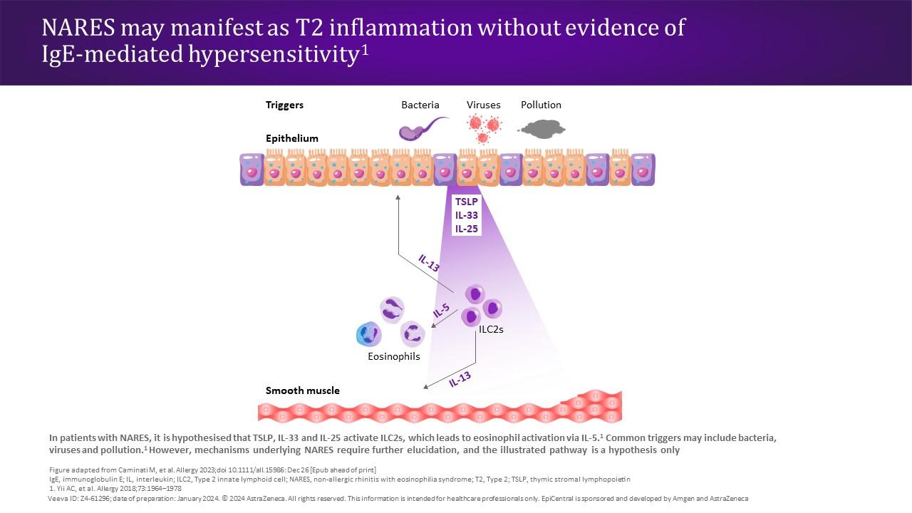
10. Yii AC, et al Allergy. 2018;73:1964–1978; 11. Adivitiya, et al Biology (Basel). 2021;10:95; 12. Zhang R, et al Int Arch Allergy Immunol. 2023;184:1–21; 13. Licari A, et al Front Pediatr. 2017;5:44; 14. Laulajainen-Hongisto A, et al Front Cell Dev Biol. 2020;8:204; 
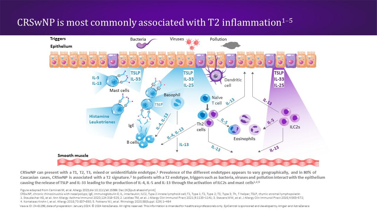
15. Stevens WW, et al J Allergy Clin Immunol Pract. 2016;4:565–572; 16. Bartemes KR, Kita H Clin Immunol. 2012;143:222–235; 
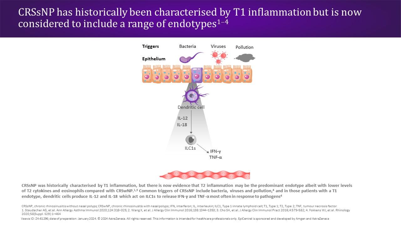
17. Fokkens WJ, et al Rhinology. 2020;58(Suppl. S29):1–464; 
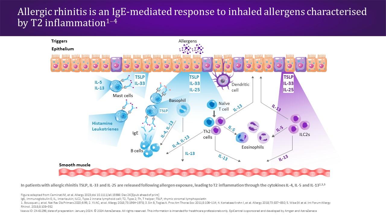
18. Bousquet J, et al Nat Rev Dis Primers. 2020;6:95; 19. Crystal RG, et al Proc Am Thorac Soc. 2008;5:772–777; 20. Davis JD, Wypych TP Mucosal Immunol. 2021;14:978–990; 21. Doeing DC, Solway J J Appl Physiol (1985). 2013;114:834–843; 22. Orlandi RR, et al Int Forum Allergy Rhinol. 2021;11:213–739; 23. Sedaghat AR, et al J Allergy Clin Immunol Pract. 2022;10:1395–1403; 24. Claeys N, et al Front Allergy. 2021;2:761388; 25. Wynne M, et al Am J Rhinol Allergy. 2019;33:782–790; 26. Zhang M, et al Int Immunopharmacol. 2023;121:110559; 27. Liao B, et al Allergy. 2015;70:1169–1180; 28. Staudacher AG, et al Ann Allergy Asthma Immunol. 2020;124:318–325; 29. Laidlaw TM, et al J Allergy Clin Immunol Pract. 2021;9:1133–1141; 30. Sehmi R Thorax. 2017;72:591–593; 31. Deng H, et al J Asthma Allergy. 2021;14:839–850; 32. Liu R, et al Front Immunol. 2021;12:530488; 33. Orlandi RR, et al Int Forum Allergy Rhinol. 2016;6(Suppl. 1):S3–S21; 34. Cho SH, et al J Allergy Clin Immunol Pract. 2016;4:575–582; 35. Bachert C, et al J Asthma Allergy. 2021;14:127–134; 36. Head K, et al Cochrane Database Syst Rev. 2016;4:CD011991; 37. Peters AT, et al Allergy Asthma Proc. 2022;43:435–445; 38. Smith KA, et al Int Forum Allergy Rhinol. 2019;9:402–408; 39. Beard S Prim Care. 2014;41:33–46; 40. Dykewicz MS, et al J Allergy Clin Immunol. 2020;146:721–767; 41. Hellings PW, et al Allergy. 2017;72:1657–1665; 42. Sin B, Togias A Proc Am Thorac Soc. 2011;8:106–114; 43. Savouré M, et al Clin Transl Allergy. 2022;12:e12130; 44. Wise SK, et al Int Forum Allergy Rhinol. 2018;8:108–352; 45. Small P, et al Allergy Asthma Clin Immunol. 2018;14(Suppl. 2):51; 46. Liu Y, et al J Immunol Res. 2022;2022:4351345; 47. Shaaban R, et al Am J Respir Crit Care Med. 2007;176:659–666; 48. Kaliner MA World Allergy Organ J. 2009;2:98–101; 49. Greiwe JC, Bernstein JA J Clin Med. 2019;8:2019; 50. Heffler E, et al Clin Exp Allergy. 2018;48:1092–1106; 51. Shamil E, Hopkins C Otolaryngol Clin North Am. 2023;56:157–168; 52. Miglani A, et al Otolaryngol Clin North Am. 2023;56:11–22; 53. Caruso C, et al Front Allergy. 2022;3:768408.