Epithelial cytokines and the inflammatory cascade

Epithelial cytokines play upstream and downstream roles in regulating immune responses in asthma.1,2

Epithelial cytokines play upstream and downstream roles in regulating immune responses in asthma1,2

  • Epithelial activation triggers the release of epithelial cytokines TSLP, IL-33 and IL-25, initiating and amplifying a cascade of complex pathways that together drive the clinical features of epithelial-driven diseases, such as asthma1–5  ​
  • Once released from the epithelium, epithelial cytokines can activate and/or modulate innate and adaptive immune responses in overlapping but distinct ways1,2​
  • Epithelial cytokines are involved in both allergic eosinophilic (T2) inflammation and beyond T2 inflammation, including structural changes5–8​
  • Multiple clinical features of asthma are associated with increased expression of TSLP, IL-33 and IL-25, including asthma severity, risk of exacerbations, reduced lung function, reduced glucocorticoid response, exaggerated ​T2 response to viral infections and potential airway remodelling8–20
  • More recently, TL1A, a novel epithelial cytokine, has also been shown to contribute to airway inflammation and remodelling through its effect on immune and structural cells21

Further understanding of the role of epithelial cytokines in the inflammatory cascade in airway disease can provide greater insights into asthma pathophysiology and possible future intervention options, with the aim of lowering disease activity through the restoration of epithelial health22,23

IL, interleukin; T2, type 2; TL1A, TNF-like cytokine 1A; TSLP, thymic stromal lymphopoietin.​
1. Roan F, et al. J Clin Invest. 2019;129:1441–1451; 2. Bartemes KR, Kita H. Clin Immunol. 2012;143:222–235; 3. McBrien CN, Menzies-Gow A. Front Med (Lausanne). 2017;4:93; 4. Varricchi G, et al. Front Immunol. 2018;9:1595; 5. Gauvreau GM, et al. Expert Opin Ther Targets. 2020;24:777–792; 6. Brusselle GG, et al. Nat Med. 2013;19:977–979; 7. Lambrecht BN, Hammad H. Nat Immunol. 2015;16:45–56; 8. Kaur D, et al. Chest. 2012;142:76–85; 9. Kato A, et al. J Immunol. 2007;179:1080–1087; 10. Beale J, et al. Sci Transl Med. 2014;6:256ra134; 11. Shikotra A, et al. J Allergy Clin Immunol. 2012;129:104–111; 12. Li Y, et al. J Immunol. 2018;200:2253–2262; 13. Ko H-K, et al. Sci Rep. 2021;11:8425; 14. Liu S, et al. J Allergy Clin Immunol. 2018;141:257–268; 15. Lee H-C, et al. J Allergy Clin Immunol. 2012;130:1187–1196; 16. Uller L, et al. Thorax. 2010;65:626–632; 17. Cao L, et al. Exp Lung Res. 2018;44:288–301; 18. Wu J, et al. Cell Biochem Funct. 2013;31:496–503; 19. Guo Z, et al. J Asthma. 2014;51:863–869; 20. Cheng D, et al. Am J Respir Crit Care Med. 2014;190:639–648; 21. Varricchi G, et al. J Allergy Clin Immunol. 2025;doi:10.1016/j.jaci.2025.02.018:Feb 24 [Epub ahead of print]; 22. Calvén J, et al. Int J Mol Sci. 2020;21:8907; 23. Duchesne M, et al. Front Immunol. 2022;13:975914.

Epithelial cytokines are rapidly released from the airway epithelium

The epithelium is a key orchestrator of the innate immune system. As described in 'The role of the epithelium in asthma', the epithelium provides a physical and immune-modulatory barrier acting as the first line of defence against environmental agents.3 

Video: Watch Professor Gianni Marone discuss the importance of epithelial cytokines in T2- and beyond-T2 inflammation in severe asthma (03:07)​

Epithelial-derived cytokines (alarmins) are the body’s ubiquitous warning signals, acting as first reactors following infection and physical or immunological insult.4 Epithelial-derived cytokines (thymic stromal lymphopoietin [TSLP], interleukin [IL]-33 and IL-25) are released by activated epithelial cells in response to injury or immunological insult.2,5  

The mechanism of epithelial-cytokine release differs from the production of traditional cytokines, which are secreted by a wide-range of immune cells in response to inflammation and infection.6 In asthma, epithelial-derived cytokines, produced by both immune and non-immune cells, are released in response to a variety of triggers present at the airway epithelium, such as pathogens, cytokines, aeroallergens, mechanical injury and air pollutants.1,2,7,8 Specifically, TSLP expression and release from epithelial cells is increased in response to a broad array of stimuli, including mechanical injury, infection, inflammatory cytokines and fungi.1,8,9 However, the activity of IL-33 is regulated both by its localisation within the cell and by proteolytic cleavage.1 Although IL-33 lacks a signal sequence required for conventional secretory pathways, it can be released as an “alarmin” in response to cellular injury or stress.10,11 

TSLP, IL-33 and IL-25 (also known as IL-17E)1 have a pleiotropic role in promoting the development of inflammation in patients with asthma by activating specific receptors on a variety of immune and non-immune cells.1 In particular, TSLP exerts its pleiotropic functions by binding to a high affinity heteromeric complex composed of TSLPR chain and IL-7R⍺.8

Image
The inflammatory cascade in asthma: role of epithelial cytokines

The inflammatory cascade in asthma: role of epithelial cytokines

Download resource

The inflammatory cascade in asthma: role of epithelial cytokines  

Once released from the epithelium, epithelial cytokines can activate and/or modulate innate and adaptive immune responses in overlapping but distinct ways.1,2 The specificity of IL-33, TSLP and IL-25 in the modulation of Type 2 (T2) inflammation is mediated by the selective expression of their different receptors on immune cells.1 While IL-33, TSLP and IL-25 can play similar roles in T2 inflammation their roles are more frequently divergent.12 

Image
Epithelial cytokines cell interactions

Epithelial cytokines cell interactions

Download resource

Several cellular targets of TSLP, IL-33 and, to a lesser extent, IL-25 have been identified, including immune and non-immune cells.1,8 The activation of these cellular targets by epithelial cytokines can cause production of several downstream cytokines (eg IL-5, IL-13 and IL-4), leading to T2 inflammation.1,5,7,8,13

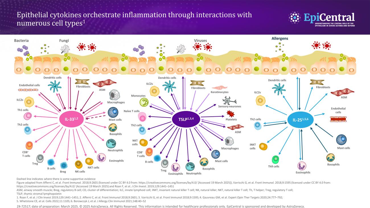
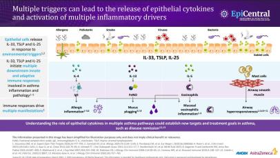
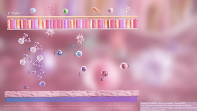
TSLP, IL-33 and, to a lesser extent, IL-25 have a large number of cellular targets.1  IL-33 targets myeloid dendritic cells, CD4+ T cells, CD8+ T cells, regulatory T cells, natural killer T cells, mast cells, macrophages, B cells, eosinophils, basophils, neutrophils, type 2 innate lymphoid cells (ILC2s), airway epithelium and fibroblasts.1 TSLP targets myeloid dendritic cells, CD4+ T cells, CD8+ T cells, regulatory T cells, natural killer T cells, B cells, mast cells, monocytes, eosinophils, basophils, ILC2s and the airway epithelium.1IL-25 targets ILC2s, CD4+ T cells, invariant natural killer T cells, airway epithelial cells and fibroblasts.1

Allergic eosinophilic (T2) inflammation, driven by allergen exposure, induces the release of epithelial cytokines (TSLP, IL-33 and IL-25), which can activate dendritic cells (DCs).5,13 Activated DCs present allergens to naïve CD4+ T cells resulting in differentiation to Th2 cells.5,13 Th2 cells, in collaboration with activated basophils, are a major source of IL-4, IL-5 and IL-13, which induce immunoglobulin (Ig)E class switching in B cells.5,13,14 These molecules activate eosinophils (predominantly driven by IL-5) and mast cells, which are key effector cells in allergic T2 inflammation.5,13,14  Click here to access the 'Epithelial cytokine inflammatory pathways' downloadable asset for a visual representation of this complex pathway.

TSLP, IL-33 and IL-25 activate ILC2s resulting in production of IL-5 and IL-13; leading to activation of eosinophils and non-allergic airway inflammation.5,13–17   

Beyond T2 inflammation, TSLP may play a role in driving structural changes through activation of fibroblasts and mast cells.5,18 In particular, human CD34+ progenitor-derived mast cells express TSLPR and IL-7R⍺.19 TSLP, in combination with certain cytokines (eg IL-1β and tumor necrosis factor [TNF]-α), causes the release of several cytokines and chemokines from mast cells.18–20 TSLP, in combination with IL-33, induces prostaglandin D2 (PGD2) production by human mast cells.21 TSLP is a survival factor for human mast cells through the activation of STAT6, providing one potential explanation for mast cell accumulation in allergic disorders.20 The structural changes mediated by mast cell and fibroblast activation ultimately lead to airway remodelling and airway hyperresponsiveness.5,22 

Further evidence suggests that TSLP, IL-33 and IL-25 may play a pivotal role beyond T2 inflammation.12 TSLP provides critical signals for T follicular helper cell (TFH) differentiation,23 human B-cell proliferation,24 and mast cell activation.18 IL-33 augments the effects of rhinovirus on the inflammatory activity of human lung vascular endothelium, which may be relevant to viral-induced asthma exacerbations.25 Mast cells, in response to IL-33, release T2 cytokines which induce upregulation of IL33 expression by epithelial cells in a feed-forward loop, suggesting that mast cells cooperate with epithelial cells through IL-33 signalling.26 IL-33 may also potentiate the release of angiogenic and lymphangiogenic factors from human mast cells.27 IL-25, which belongs to the IL-17 cytokine family, exerts its biological effects by interacting with a dimeric complex consisting of the two receptor subunits IL-17R⍺ and IL-17RB.1 IL-25 exerts a pathogenic role in allergic asthma and virus-induced exacerbations.28

Epithelial cytokines are associated with clinical features of asthma 

Multiple clinical features of asthma are associated with increased expression of TSLP and/or IL-33, including: 

  • Asthma severity29,30 
  • Risk of asthma exacerbations31  
  • Reduced lung function30 
  • Reduced glucocorticoid response32 
  • Exaggerated T2 response to viral infections9,33,34 
  • Potential airway remodelling35–37 

Additionally, increased expression of IL-25 is associated with potential airway remodelling,38 and exaggerated T2 response to viral infections.28

Click here to access more about the potential roles of TSLP, IL-33 and IL-25 in each of these clinical features.

Image
Multiple inflammatory responses in asthma inforaphic

Multiple clinical features of asthma are associated with increased expression of epithelial cytokines

Download resource

Find out more about the EpiCreator – Professor Gianni Marone

RELATED RESOURCES

Like this page? We have a host of other resources available in the ‘Scientific and Resource Library’ where you can find out more.

References

1. Roan F, et al. J Clin Invest. 2019;129:1441–1451; 2. Bartemes KR, Kita H. Clin Immunol. 2012;143:222–235; 3. Holgate ST. Immunol Rev. 2011;242:205–219; 4. Yang D, et al. Immunol Rev. 2017;280:41–56; 5. Gauvreau GM, et al. Expert Opin Ther Targets. 2020;24:777–792; 6. Lacy P, Stow JL. Blood. 2011;118:9–18; 7. McBrien CN, Menzies-Gow A. Front Med (Lausanne). 2017;4:93; 8. Varricchi G, et al. Front Immunol. 2018;9:1595; 9. Kato A, et al. J Immunol. 2007;179:1080–1087; 10. Kouzaki H, et al. J Immunol. 2011;186:4375–4387; 11. Kakkar R, et al. J Biol Chem. 2012;287:6941–6948; 12. Porsbjerg CM, et al. Eur Respir J. 2020;56:2000260; 13. Brusselle GG, et al. Nat Med. 2013;19:977–979; 14. Lambrecht BN, Hammad H. Nat Immunol. 2015;16:45–56; 15. Brusselle G, Bracke K. Ann Am Thorac Soc. 2014;11(Suppl. 5):S322–S328; 16. Halim TYF, et al. Immunity. 2012;36:451–463; 17. Martin NT, Martin MU. Nat Immunol. 2016;17:122–131; 18. Kaur D, et al. Chest. 2012;142:76–85; 19. Allakhverdi Z, et al. J Exp Med. 2007;204:253–258; 20. Han N-R, et al. J Invest Dermatol. 2014;134:2521–2530; 21. Buchheit KM, et al. J Allergy Clin Immunol. 2016;137:1566–1576; 22. Ishmael FT. J Am Osteopath Assoc. 2011;111:S11–S17; 23. Pattarini L, et al. J Exp Med. 2017;214:1529–1546; 24. Milford T-AM, et al. Eur J Immunol. 2016;46:2155–2161; 25. Gajewski A, et al. Allergy. 2021;76:2282–2285; 26. Altman MC, et al. J Clin Invest. 2019;129:4979–4991; 27. Cristinziano L, et al. Cells. 2021;10:145; 28. Beale J, et al. Sci Transl Med. 2014;6:256ra134; 29. Shikotra A, et al. J Allergy Clin Immunol. 2012;129:104–111; 30. Li Y, et al. J Immunol. 2018;200:2253–2262; 31. Ko H-K, et al. Sci Rep. 2021;11:8425; 32. Liu S, et al. J Allergy Clin Immunol. 2018;141:257–268; 33. Lee H-C, et al. J Allergy Clin Immunol. 2012;130:1187–1196; 34. Uller L, et al. Thorax. 2010;65:626–632; 35. Cao L, et al. Exp Lung Res. 2018;44:288–301; 36. Wu J, et al. Cell Biochem Funct. 2013;31:496–503; 37. Guo Z, et al. J Asthma. 2014;51:863–869; 38. Cheng D, et al. Am J Respir Crit Care Med. 2014;190:639–648.