Complexity of severe asthma

Inflammation in asthma is complex and heterogeneous, which makes it challenging to manage.1–4

Severe asthma is characterised by complex, heterogeneous and dynamic airway inflammation1–4

  • Activation of the airway epithelium, following exposure to environmental triggers, results in epithelial cytokine release1,5​
  • This release of epithelial cytokines can lead to initiation of multiple downstream inflammatory pathways, including T2 inflammation (allergic and/or eosinophilic) and beyond T2 inflammation (structural changes to the airway)1,5–8​
  • Patients may display overlapping, dynamic inflammatory pathways, which can change over time owing to different circumstances;1–4,9 many patients are thought to have multiple drivers of disease2
  • Dynamic, objective and diagnostic clinical biomarkers can help to identify phenotypes and endotypes of severe asthma and serve as valuable tools to support in the selection of an appropriate treatment and improving patient outcomes10

Improved understanding of the key inflammatory pathways and mechanisms that underpin epithelial-driven diseases will continue to direct phenotypic research, with the goal of finding ways to restore epithelial health, lower disease activity, and ultimately achieve clinical remission in asthma8,11–14

T2, type 2​
1. Busse WW. Allergol Int. 2019;68:158–166; 2. Tran TN, et al. Ann Allergy Asthma Immunol. 2016;116:37–42; 3. Price D, Canonica GW. World Allergy Organ J. 2020;13:100380; 4. Kupczyk M, et al. Allergy. 2014;69:1198–1204; 5. Lambrecht BN, Hammad H. Nat Med. 2012;18:684–692; 6. Cao L, et al. Exp Lung Res. 2018;44:288–301; 7. Wu J, et al. Cell Biochem Funct. 2013;31:496–503; 8. Gauvreau GM, et al. Expert Opin Ther Targets. 2020;24:777–792; 9. Kuruvilla ME, et al. Clin Rev Allergy Immunol. 2019;56:219–233; 10. Carr TF, Kraft M. Ann Allergy Asthma Immunol. 2018;121:414–420; 11. Kaur R, Chupp G. J Allergy Clin Immunol. 2019;144:1–12; 12. Agache I, et al. Allergy. 2012;67:835–846. 13. Hiemstra PS, et al. Eur Respir J. 2015;45:1150–1162; 14. Carpaij OA, et al. Pharmacol Ther. 2019;201:8–24.

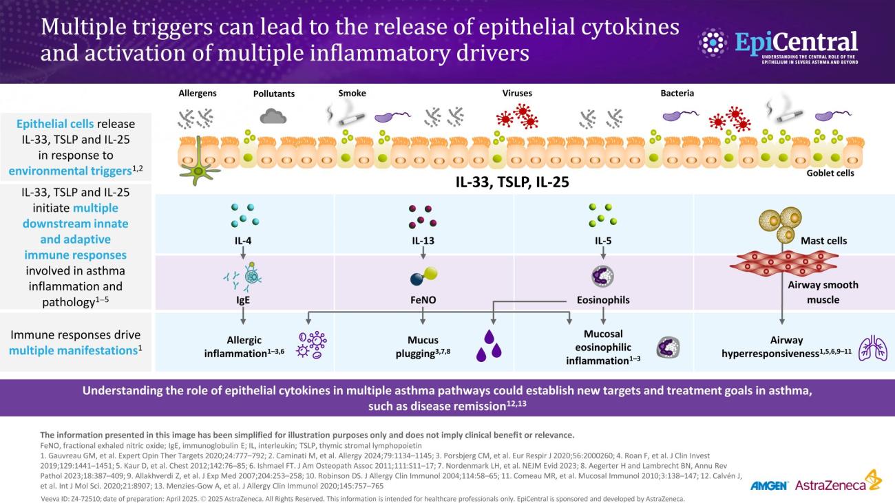
Inflammation in severe asthma

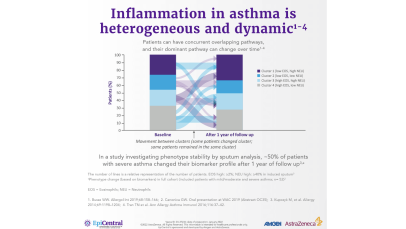
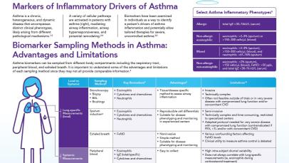
Asthma-associated inflammation is complex and heterogeneous,1–4 and numerous cell types, mediators and downstream immune pathways are involved.1,3–6 Multiple inflammatory endotypes have been characterised – allergic and eosinophilic inflammation, to name a few.5 

Video: Watch Professor Christopher Brightling introduce the complexity of severe asthma ​ (04:09)

Overlapping inflammatory pathways in severe asthma 

Activation of the airway epithelium by exposure to environmental triggers results in the production of epithelial cytokines and leads to a cascade of events that result in airway inflammation and the clinical manifestations of asthma.1,7 This airway inflammation, alongside airway remodelling, drives changes in asthma pathophysiology and leads to airway hyperresponsiveness and airflow obstruction.1 

The production of epithelial cytokines leads to multiple downstream immune pathways in patients with severe asthma, including:1 

  • Type 2 (T2) inflammation, typically marked by allergic and/or eosinophilic inflammation1 
  • Mechanisms beyond T2 inflammation, characterised by neutrophilic cellular infiltrate or paucigranulocytic inflammation, and airway hyperresponsiveness, airway remodelling or microbial dysbiosis, which may exist together with T2 inflammation1,8,9 

While many patients have T2 disease,1,2 a sizeable group has mechanisms that are beyond T2 disease.2  

Video: Watch Professor Ian Pavord explain asthma heterogeneity and the dynamic nature of airway inflammation in asthma (01:05)

Up to 60% of patients may have multiple drivers of inflammation;1–4,10 as seen in the International Severe Asthma Registry (ISAR), >50% of patients had two or more elevated biomarkers.10 Therefore, it is likely that many patients have a mixture of pathways that drive their disease.1,2

The dominant pathway may change over time owing to different circumstances, such as changes in medications, treatment adherence, exacerbations or exposure to allergens.1–4 In a biomarker study conducted in patients with severe asthma, the phenotype changed in ~50% of patients based on their sputum biomarker clustering after 1 year of follow-up.4  

Upstream activation of epithelial cytokines can lead to initiation of multiple downstream inflammatory pathways.11 The wide spectrum and overlap of downstream pathways in severe asthma mean that diagnosis and treatment can be challenging.2 For example, in one US study, around a third of adults with severe asthma were classified as having both an allergic and an eosinophilic (blood eosinophil cut-off was 300 cells/µL) phenotype.2 It would be advantageous to further understand any overlap and common drivers (such as epithelial cytokines) of the downstream endotypes of severe asthma.12,13 Understanding this may ensure appropriate treatments are chosen to address the active inflammatory pathways at the source, ultimately leading to improved patient care and restoration of epithelial health.6,12,14

Biomarkers in severe asthma  

Dynamic, objective and diagnostic biomarkers can help to identify phenotypes and endotypes of severe asthma and help in the selection of an appropriate treatment.15 As such, it is important that clinicians are able to interpret biomarkers effectively to improve patient outcomes.15  

Various biomarkers of T2-mediated inflammation, including specific blood immunoglobulin (Ig) E, blood or sputum eosinophils, and fractional exhaled nitric oxide (FeNO), are available to clinicians1,15,16 and these, among others, can be used in clinical practice for phenotyping of severe asthma.15 

  • Allergen-specific blood IgE levels, measured via a blood or skin prick testing, are higher in patients with allergic asthma compared with healthy individuals and can be used as a surrogate measure for atopy15,17 
  • Blood eosinophils and sputum eosinophils are surrogate markers of T2-inflammation and the T2-inflammatory cytokine, interleukin (IL)-5, which is required for eosinophil activation and survival.16 These are useful biomarkers as patients with higher eosinophil counts are prone to experiencing severe disease and poorer asthma outcomes than patients with lower eosinophil counts18 
  • FeNO is a biomarker of airway epithelial cell exposure to IL-13, IL-4 and IL-5.11,16 These cytokines upregulate inducible nitric oxide synthase (iNOS) in the airway epithelium, and result in increased nitric oxide production.16 A high FeNO measurement correlates with airway eosinophilia in asthma and indicates increased airway T2 inflammation16

Biomarker assessment in a 32-year-old male patient with severe asthma

Image
Module 2_Inflammatory cascade

Biomarker assessment in a 32-year-old male patient with severe asthma

Download resource

Image
Phenotyping a patient with severe asthma

Phenotyping a patient with severe asthma

Download resource

It is important to note that biomarker levels may be affected by treatments.17 Biomarkers of T2 inflammation are often suppressed by inhaled corticosteroids and oral corticosteroids; therefore, eosinophils and FeNO assessments are encouraged before commencing a short course or maintenance OCS, or on the lowest possible OCS dose.17  

Some gaps exist in the clinical predictive value of existing biomarkers owing to the challenge of identifying a single predominant endotype of severe asthma.6 Furthermore, there are currently no readily available biomarkers in clinical practice that identify T2-independent asthma.11,19,20 For now, biomarker data need to be interpreted alongside symptoms and lung function, and need to focus on identifying tractable features of asthma, such as airway hyperresponsiveness.21 

Video: Professor Christopher Brightling explains the use of biomarkers in airway inflammation and their predictive value (07:11)

Find out more about the EpiCreator – Professor Christopher Brightling

RELATED RESOURCES

Like this page? We have a host of other resources available in the ‘Scientific and Resource Library’ where you can find out more.

References
1. Busse WW. Allergol Int. 2019;68:158–166; 2. Tran TN, et al. Ann Allergy Asthma Immunol. 2016;116:37–42; 3. Price D, Canonica GW. World Allergy Organ J. 2020;13:100380 (Abstract OC35); 4. Kupczyk M, et al. Allergy. 2014;69:1198–1204; 5. Barnes PJ. Pathophysiology of asthma. In: European Respiratory Society Monograph. 2003:84–113; 6. Gauvreau GM, et al. Expert Opin Ther Targets. 2020;24:777–792; 7. Lambrecht BN, Hammad H. Nat Med. 2012;18:684–692; 8. Cao L, et al. Exp Lung Res. 2018;44:288–301; 9. Wu J, et al. Cell Biochem Funct. 2013;31:496–503; 10. Denton E, et al. J Allergy Clin Immunol Pract. 2021;9:2680–2688; 11. Kuruvilla ME, et al. Clin Rev Allergy Immunol. 2019;56:219–233; 12. Kaur R, Chupp G. J Allergy Clin Immunol. 2019;144:1–12; 13. Agache I, et al. Allergy. 2012;67:835–846; 14. Russell RJ, et al. Eur Respir J. 2024;63:2301397; 15. Carr TF, Kraft M. Ann Allergy Asthma Immunol. 2018;121:414–420; 16. Peters MC, et al. Curr Allergy Asthma Rep. 2016;16:71; 17. Global Initiative for Asthma (GINA). Global strategy for asthma management and prevention. 2024. Available from: https://ginasthma.org/wp-content/uploads/2024/05/GINA-2024-Strategy-Report-24_05_22_WMS.pdf (Accessed 24 April 2025); 18. Kostikas K, et al. Curr Drug Targets. 2018;19:1882–1896; 19. Schleich F, et al. Curr Top Med Chem. 2016;16:1561–1573; 20. Quoc QL, et al. Exp Mol Med. 2021;53:1170–1179; 21. James A, Hedlin G. Curr Treat Options Allergy. 2016;3:439–452.