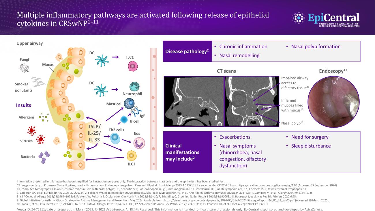
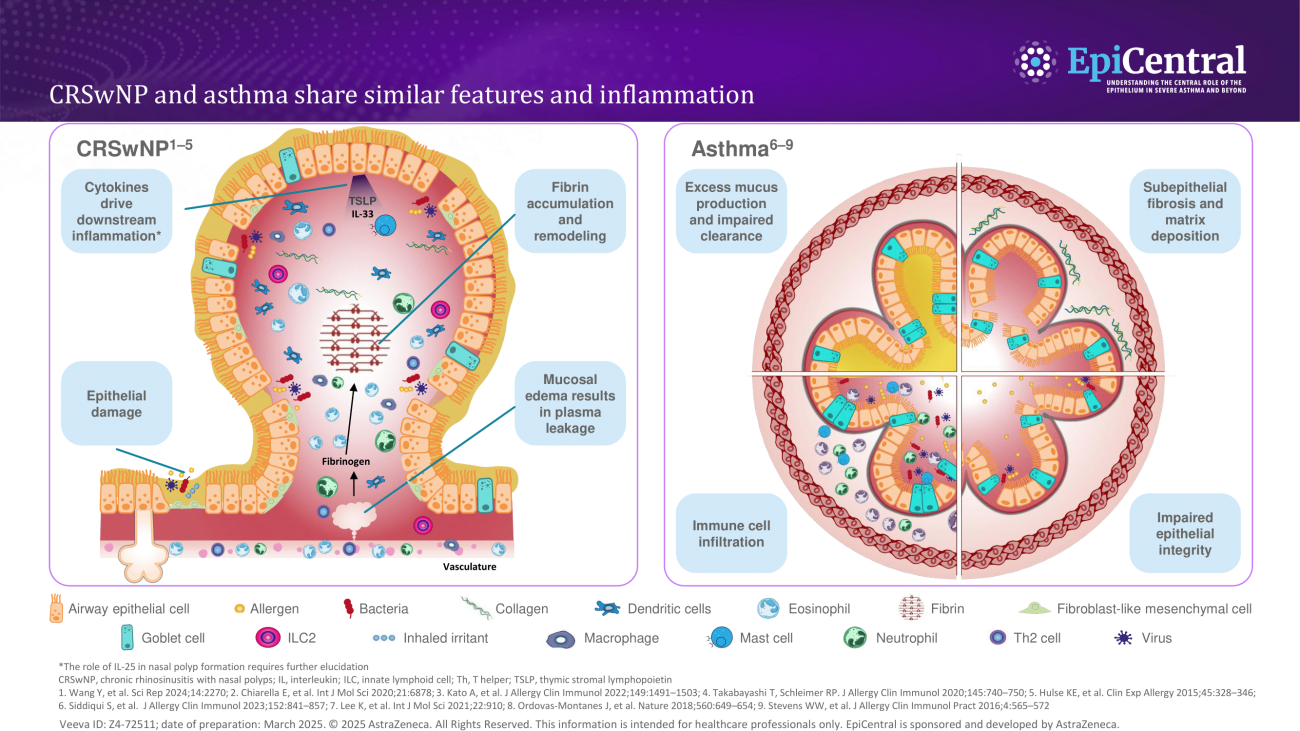
Chronic rhinosinusitis and the central role of the nasal epithelium

Epithelial dysregulation plays a central role in the pathogenesis of chronic rhinosinusitis.1

Module synopsis

  • Chronic rhinosinusitis (CRS) is characterised by chronic sinonasal inflammation and divided into two main phenotypes: CRS with nasal polyps (CRSwNP) and CRS without nasal polyps (CRSsNP)2
  • Estimates of CRS prevalence range from less than 5% of the population to more than 10%3 
  • The condition has a significant psychological and social burden, but patients perceive an underestimation of disease burden by healthcare professionals (HCPs)4 
  • Epithelial dysfunction is an important characteristic of CRS and impedes the ability of the epithelium to act as a physical and immune barrier against the external environment1 
  • Elevated expression of epithelial cytokines has been demonstrated in the nasal epithelium of patients with CRSwNP,5 and levels of thymic stromal lymphopoietin (TSLP), the TSLP receptor (TSLPR) and the interleukin (IL)-33 receptor (ST2L) have been shown to correlate with markers of disease severity in eosinophilic CRSwNP6 

1. Wynne M, et al. Am J Rhinol Allergy. 2019;33:782–790; 2. Orlandi RR, et al. Int Forum Allergy Rhinol. 2021;11:213–739; 3. Sedaghat AR, et al. J Allergy Clin Immunol Pract. 2022;10:1395–1403; 4. Claeys N, et al. Front Allergy. 2021;2:761388; 5. Zhang M, et al. Int Immunopharmacol. 2023;121:110559; 6. Liao B, et al. Allergy. 2015;70:1169–1180.

Professor Claire Hopkins' and Dr Anju Peters' thoughts on chronic rhinosinusitis and the central role of the nasal epithelium

Image
Peters and Hopkins quote

Insights from our EpiCollaborators

Download resource

What is CRS?  

Chronic rhinosinusitis (CRS) is divided into two main phenotypes: CRS with nasal polyps (CRSwNP) and CRS without nasal polyps (CRSsNP).2 There are other phenotypes of CRS, including aspirin-exacerbated respiratory disease (AERD), infectious rhinosinusitis and fungal rhinosinusitis, a subtype of which is allergic fungal rhinosinusitis (AFRS).7

Estimates of CRS prevalence vary: over 10% of the population has been estimated to have CRS based on the presence of either symptoms or objective evidence alone, while the presence of both has produced estimates of less than 5%.3 Reported prevalence also varies between countries.8

Based on a retrospective dataset from patients with a history of CRS with positive sinus CT, evaluated in a US clinic (2002–2012), CRSsNP accounts for approximately 80% of cases, while patients with CRSwNP, although constituting a smaller proportion of CRS cases, tend to have more severe clinical disease.9

CRS is diagnosed using subjective and objective evidence of chronic sinonasal inflammation.2 In terms of the former, the condition is characterised by ≥12 weeks of two or more of the following symptoms: nasal obstruction or congestion, nasal discharge, hyposmia, and/or facial pressure or pain.2,8 In terms of the latter, objective criteria include evidence of inflammation via nasal endoscopy or computed tomography (CT).2

The burden of disease

CRS has a substantial negative impact on health-related quality of life.4,10 Patients report that symptoms have a major impact on daily activities, including reducing food enjoyment, reducing sleep quality and causing feelings of embarrassment; thus, the disease represents a significant psychological and social burden.4 

In fact, the symptoms of CRS are perceived to result in a comparable health state to those of Parkinson’s disease or congestive heart failure, as defined by mean Short-Form Six-Dimension (SF-6D) impairment scores.10 Furthermore, compared with non-AERD CRSwNP or CRSsNP, data suggest that patients with AERD suffer the most burdensome symptoms.11

Additionally, several studies have shown an association between CRS and increased rates of depression and anxiety.12,13

Despite the significant impact of CRS, patients perceive an underestimation of disease burden by healthcare professionals (HCPs).4 Timely diagnosis and management is critical for patient outcomes and effective healthcare utilisation; delayed surgical intervention for CRS is associated with greater postoperative healthcare needs.14,15 Yet, patients’ feelings of being dismissed can act as a barrier to effective communication with HCPs, which contributes to delays in diagnosis and, therefore, treatment.16

The unmet need in CRS

Current treatment algorithms for CRS recommend intranasal corticosteroids and/or saline rinses as the first-line treatment and as maintenance therapy.17 Subsequent treatment is advanced stepwise, through short courses of oral corticosteroids (OCS), endoscopic sinus surgery (ESS), and then revision surgery or, more recently, approved biologics.8,17

However, there are limitations to these approaches: OCS often provide only temporary alleviation of symptoms such as anosmia,18 and recurrent use of systemic corticosteroids (SCS) is associated with an increased risk of several adverse events, including pneumonia, osteoporosis, cardiovascular disease, cataracts, sleep apnoea, depression/anxiety and diabetes.19

Even appropriate medical therapy and surgery often do not achieve disease control in the long term.20 Based on an observational study in Belgium (2013–2014) in adult patients with CRS (n=560), at least 40% of patients may have uncontrolled disease 3–5 years after functional ESS.21 In a prospective multi-centre cohort in North America from 2004–2015 of adult patients undergoing ESS for medically recalcitrant CRSwNP, 35% (n=68) of patients with CRSwNP who underwent ESS were found to have nasal polyp recurrence at 6-month follow-up.22

  • Although >90% of patients are prescribed intranasal corticosteroids,23 they often provide insufficient control24
  • In a survey from Italy, ~70% of physicians (n=437) reported that OCS provide only temporary symptom relief, leading to recurrent OCS courses18
  • In a US real-world retrospective study of patients with CRSwNP receiving biologics (n=2208), ~30% required OCS two more times in 12 months25
  • Based on an observational study in Belgium from 2013–2014 in adult patients with CRS (n=560), 80% of patients reported inadequately controlled symptoms within 3 to 5 years after surgery. In those who reported uncontrolled symptoms, blocked nose and smell disorders were also reported21

Pathophysiology: the central role of the epithelium

Advances in understanding have enabled a paradigm shift from viewing CRS pathophysiology through the lens of airflow obstruction to a model in which the airway epithelium plays a crucial role.26

The airway and nasal epithelia orchestrate the complex interactions between the body’s external and internal environments.27,28 The nasal passage is the first point of entry for environmental triggers (such as pathogens, allergens and pollutants) and odorants entering the respiratory tract that interact with the highly specialised nasal and olfactory epithelia, respectively.28–30

The healthy nasal epithelium maintains homeostasis through a variety of immune and non-immune functions including acting as a physical barrier to environmental agents, driving mucociliary clearance, orchestrating innate immune responses through pattern-recognition receptors and regulating adaptive immune cells via the release of cytokines.28

Image
The epithelium plays a critical role in CRSwNP

The epithelium plays a critical role in CRSwNP

Download resource

However, dysregulation of the epithelium is an important characteristic of CRS whereby the ability of the epithelium to act as a robust physical and immune barrier against the external environment becomes impeded:1 

  • There is excess secretion and altered composition of mucus, mainly associated with glandular hyperplasia in CRSsNP and goblet cell hyperplasia in CRSwNP31 
  • Reduced ciliary beating, in conjunction with excess mucus, impairs mucociliary clearance, contributing to chronic infection and inflammation28,32 
  • The olfactory epithelium also appears to be altered in CRS.33 Olfactory dysfunction is a key predictor of reduced quality of life in patients with CRSwNP.34 In part, anosmia/hyposmia may be caused by reduced airflow due to physical obstruction by nasal polyps.33 However, there is also evidence that inflammation within the olfactory epithelium leads to the impairment or death of sensory neurons, and hence the loss of sense of smell33
  • Recent evidence indicates that damage to the epithelium may initiate nasal polyp formation through the process of epithelial-mesenchymal transition (EMT).35 In response to damage, epithelial cells can proliferate and undergo EMT, transforming into fibroblasts that promote tissue remodelling and, ultimately, polyp formation35 

Image
Nasal polyp formation is an epithelial-driven process

Nasal polyp formation is an epithelial-driven process

Download resource

Epithelial barrier dysfunction and impaired mucociliary clearance can lead to accumulation of pathogens and allergens, amplifying the inflammatory immune responses.29 Inflammation and remodelling at the epithelium promote one another in a positive feedback loop, exacerbating CRS pathology.36 Restoration of epithelial homeostasis may play a role in achieving disease remission in CRSwNP.37

Pathophysiology: the endotypes of CRS 

Understanding the mechanisms of pathogenesis is critical to providing optimised patient care, but this is challenging because of the highly heterogeneous nature of CRS.28,38

It was previously thought that CRSsNP and CRSwNP were characterised by distinct inflammatory endotypes, the former by Type (T)1 inflammation and the latter by T2 inflammation.28 However, recent studies show that both phenotypes can manifest in three main endotypes: T1, T2 or T3 inflammation.28,38 These endotypes are characterised by their profile of elevated cytokines and immune cells.38

Image
Chronic rhinosinusitis is a heterogenous disease with three main inflammatory endotypes

Chronic rhinosinusitis is a heterogenous disease with three main inflammatory endotypes

Download resource

Adding additional layers of complexity, a number of patients present with a mixed endotype or no clear endotype,39 and endotype prevalence appears to vary geographically:

  • T2 is the predominant endotype of CRSwNP and CRSsNP in Western countries.39 This predominance is weaker in the CRSsNP population, and a higher proportion of this patient population have no clear endotype39 
  • Asian countries show higher proportions of T1 and T3 endotypes of CRSwNP.40,41 However, there has been an increasing prevalence of T2 inflammation in the past two decades, possibly owing to shifting environmental factors40,41

Pathophysiology: the central role of epithelial cytokines

The epithelium can regulate various mediators associated with the endotypes of CRS, such as T2 cytokines, through the release of the epithelial cytokines thymic stromal lymphopoietin (TSLP), interleukin (IL)-25 and IL-33.42,43 These cytokines play key roles in the initiation of inflammatory pathways and structural changes underlying respiratory diseases such as asthma.44,45

Elevated expression of TSLP, IL-33 and IL-25 has also been demonstrated in the nasal epithelium of patients with CRSwNP.5 Levels of TSLP, the TSLP receptor (TSLPR) and the IL-33 receptor (ST2L) have been shown to correlate not only with markers of T2 inflammation in CRSwNP, but also with markers of disease severity in eosinophilic CRSwNP.6

Image
Inflammatory cascade in CRSwNP

Inflammatory pathways in CRSwNP

Download resource

Further investigation of IL-25 expression in CRSwNP is required.28 There are conflicting results about cytokine levels in CRSsNP, and further research with larger sample sizes is required.46

Comorbidities and ‘united airways disease’ 

CRS and asthma are both associated with epithelial barrier dysfunction and shared profiles of inflammatory cytokines.47

This shared pathogenesis is evidence towards the ‘united airways disease’ concept, which describes how inflammatory diseases of the upper and lower airways frequently co-occur and share similar pathophysiology.47

In regard to frequent comorbidity, estimates suggest that up to 56% of patients with CRSwNP have comorbid asthma,23 and asthma severity is positively correlated with CRS severity (as measured by Lund-Mackay scores).48 Similarly, in patients with asthma, CRS is associated with increased asthma exacerbation frequency49 and is an independent negative predictor of quality of life.50

Image
CRSwNP and asthma share similar features and inflammation

CRSwNP and asthma share similar features and inflammation

Download resource

A number of other comorbidities, such as allergy and chronic obstructive pulmonary disease (COPD), are more frequent in patients with CRS than in controls.51

The content for this module was created with the support of Professor Claire Hopkins and Dr Anju Peters.

RELATED RESOURCES

Like this page? We have a host of other resources available in the ‘Scientific and Resource Library’ where you can find out more.

References

1. Wynne M, et al. Am J Rhinol Allergy. 2019;33:782–790; 2. Orlandi RR, et al. Int Forum Allergy Rhinol. 2021;11:213–739; 3. Sedaghat AR, et al. J Allergy Clin Immunol Pract. 2022;10:1395–1403; 4. Claeys N, et al. Front Allergy. 2021;2:761388; 5. Zhang M, et al. Int Immunopharmacol. 2023;121:110559; 6. Liao B, et al. Allergy. 2015;70:1169–1180;
7. Cho SH, et al. J Allergy Clin Immunol Pract. 2020;8:1505–1511; 8. Fokkens WJ, et al. Rhinology. 2020;58(Suppl. S29):1–464; 9. Benjamin MR, et al. J Allergy Clin Immunol Pract. 2019;7:1010–1016; 10. Soler ZM, et al. Laryngoscope. 2011;121:2672–2678; 11. Schneider S, et al. J Clin Med. 2020;9:925; 12. Kim J-Y, et al. JAMA Otolaryngol Head Neck Surg. 2019;145:313–319; 13. Tomoum MO, et al. Int Forum Allergy Rhinol. 2015;5:674–681; 14. Hopkins C, et al. Rhinology. 2015;53:18–24; 15. Hopkins C, et al. Rhinology. 2015;53:10–17; 16. Vennik J, et al. BMJ Open. 2019;9:e022644; 17. Hellings PW, et al. Rhinology. 2023;61:85–89; 18. De Corso E, et al. J Pers Med. 2022;12:897; 19. Price DB, et al. J Asthma Allergy. 2018;11:193–204; 20. Fokkens WJ, et al. Allergy. 2019;74:2312–2319; 21. van der Veen J, et al. Allergy. 2017;72:282–290; 22. DeConde AS, et al. Laryngoscope. 2017;127:550–555; 23. Chen S, et al. Curr Med Res Opin. 2020;36:1897–1911; 24. Starry A, et al. Allergy. 2022;77:2725–2736; 25. Peters A, et al. J Ann Allergy Asthma Immunol. 2024;133(Suppl.):S8 (Abstract D020); 26. Yan B, et al. J Allergy Clin Immunol. 2024;153:1206–1214; 27. Vroling AB, et al. Allergy. 2008;63:1110–1123; 28. Kato A, et al. J Allergy Clin Immunol. 2022;149:1491–1503; 29. Ha J-G, Cho H-J. Int J Mol Sci. 2023;24:14229; 30. Harkema JR, et al. Toxicol Pathol. 2006;34:252–269; 31. Tu Y, et al. Inflammation. 2021;44:1937–1948; 32. Gudis D, et al. Am J Rhinol Allergy. 2012;26:1–6; 33. Yan X, et al. Laryngoscope Investig Otolaryngol. 2020;5:992–1002; 34. Mullol J, et al. J Allergy Clin Immunol Pract. 2022;10:1434–1453; 35. Wang Y, et al. Sci Rep. 2024;14:2270; 36. Gong X, et al. Front Immunol. 2023;14:1238673; 37. Chan Y, et al. J Otolaryngol Head Neck Surg. 2023;52:50; 38. Staudacher AG, et al. Ann Allergy Asthma Immunol. 2020;124:318–325; 39. Stevens WW, et al. J Allergy Clin Immunol Pract. 2019;7:2812–2820; 40. Zhang Y, et al. J Allergy Clin Immunol. 2017;140:1230–1239; 41. Ryu G, et al. Precis Future Med. 2022;6:170–176; 42. Schleimer RP. Annu Rev Pathol. 2017;12:331–357; 43. Laidlaw TM, et al. J Allergy Clin Immunol Pract. 2021;9:1133–1141; 44. Gauvreau GM, et al. Allergy. 2023;78:402–417; 45. Duchesne M, et al. Front Immunol. 2022;13:975914; 46. Cho SH, et al. J Allergy Clin Immunol Pract. 2016;4:575–582; 47. Fokkens W, Reitsma S. Otolaryngol Clin North Am. 2023;56:1–10; 48. Lin DC, et al. Am J Rhinol Allergy. 2011;25:205–208; 49. Denlinger LC, et al. Am J Respir Crit Care Med. 2017;195:302–313; 50. Ek A, et al. Allergy. 2013;68:1314–1321; 51. Khan A, et al. Rhinology. 2019;57:32–42.